Filter
Associated Lab
Publication Date
- November 15, 2008 (1) Apply November 15, 2008 filter
- November 14, 2008 (1) Apply November 14, 2008 filter
- November 12, 2008 (2) Apply November 12, 2008 filter
- November 7, 2008 (2) Apply November 7, 2008 filter
- November 1, 2008 (2) Apply November 1, 2008 filter
- Remove November 2008 filter November 2008
- Remove 2008 filter 2008
Type of Publication
8 Publications
Showing 1-8 of 8 resultsA derivative of rhodamine 110 has been designed and assessed as a probe for cytochrome P450 activity. This probe is the first to utilize a ’trimethyl lock’ that is triggered by cleavage of an ether bond. In vitro, fluorescence was manifested by the CYP1A1 isozyme with k(cat)/K(M)=8.8x10(3)M(-1)s(-1) and K(M)=0.09microM. In cellulo, the probe revealed the induction of cytochrome P450 activity by the carcinogen 2,3,7,8-tetrachlorodibenzo-p-dioxin, and its repression by the chemoprotectant resveratrol.
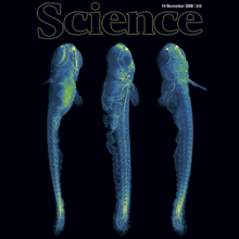
A long-standing goal of biology is to map the behavior of all cells during vertebrate embryogenesis. We developed digital scanned laser light sheet fluorescence microscopy and recorded nuclei localization and movement in entire wild-type and mutant zebrafish embryos over the first 24 hours of development. Multiview in vivo imaging at 1.5 billion voxels per minute provides "digital embryos," that is, comprehensive databases of cell positions, divisions, and migratory tracks. Our analysis of global cell division patterns reveals a maternally defined initial morphodynamic symmetry break, which identifies the embryonic body axis. We further derive a model of germ layer formation and show that the mesendoderm forms from one-third of the embryo’s cells in a single event. Our digital embryos, with 55 million nucleus entries, are provided as a resource.
At excitatory synapses, decreases in cleft [Ca] arising from activity-dependent transmembrane Ca flux reduce the probability of subsequent transmitter release. Intense neural activity, induced by physiological and pathological stimuli, disturb the external microenvironment reducing extracellular [Ca] ([Ca](o)) and thus may impair neurotransmission. Increases in [Ca](o) activate the extracellular calcium sensing receptor (CaSR) which in turn inhibits nonselective cation channels at the majority of cortical nerve terminals. This pathway may modulate synaptic transmission by attenuating the impact of decreases in [Ca](o) on synaptic transmission. Using patch-clamp recording from isolated cortical terminals, cortical neuronal pairs and isolated neuronal soma we examined the modulation of synaptic transmission by CaSR. EPSCs were increased on average by 88% in reduced affinity CaSR-mutant (CaSR(-/-)) neurons compared with wild-type. Variance-mean analysis indicates that the enhanced synaptic transmission was due largely to an increase in average probability of release (0.27 vs 0.46 for wild-type vs CaSR(-/-) pairs) with little change in quantal size (23 +/- 4 pA vs 22 +/- 4 pA) or number of release sites (11 vs 13). In addition, the CaSR agonist spermidine reduced synaptic transmission and increased paired-pulse depression at physiological [Ca](o). Spermidine did not affect quantal size, consistent with a presynaptic mechanism of action, nor did it affect voltage-activated Ca channel currents. In summary, reduced CaSR function enhanced synaptic transmission and CaSR stimulation had the opposite effect. Thus CaSR provides a mechanism that may compensate for the fall in release probability that accompanies decreases in [Ca](o).
The chemical senses, smell and taste, are the most poorly understood sensory modalities. In recent years, however, the field of chemosensation has benefited from new methods and technical innovations that have accelerated the rate of scientific progress. For example, enormous advances have been made in identifying olfactory and gustatory receptor genes and mapping their expression patterns. Genetic tools now permit us to monitor and control neural activity in vivo with unprecedented precision. New imaging techniques allow us to watch neural activity patterns unfold in real time. Finally, improved hardware and software enable multineuron electrophysiological recordings on an expanded scale. These innovations have enabled some fresh approaches to classic problems in chemosensation.
Landmark correspondences can be used for various tasks in image processing such as image alignment, reconstruction of panoramic photographs, object recognition and simultaneous localization and mapping for mobile robots. The computer vision community knows several techniques for extracting and pairwise associating such landmarks using distinctive invariant local image features. Two very successful methods are the Scale Invariant Feature Transform (SIFT)1 and Multi-Scale Oriented Patches (MOPS).2
We implemented these methods in the Java programming language3 for seamless use in ImageJ.4 We use it for fully automatic registration of gigantic serial section Transmission Electron Microscopy (TEM) mosaics. Using automatically detected landmark correspondences, the registration of large image mosaics simplifies to globally minimizing the displacement of corresponding points.
We present here an introduction to automatic landmark correspondence detection and demonstrate our implementation for ImageJ. We demonstrate the application of the plug-in on diverse image data.
In order to study anatomy of organisms with high-resolution there is an increasing demand to image large specimen in three dimensions (3D). Confocal microscopy is able to produce high-resolution 3D images, but these are limited by its relatively small field of view compared to the size of large biological specimens. To overcome this drawback, motorized stages moving the sample are used to create a tiled scan of the whole specimen. The physical coordinates provided by the microscope stage are not precise enough to allow reconstruction (”Stitching”) of the whole image from individual image stacks.
We developed an algorithm, as well as an ImageJ plug-in, based on the Fourier Shift Theorem that computes all possible translations (x, y, z) between two 3D images at once, yielding the best overlap in terms of the cross correlation measure. Apart from the obvious gain in computation time it has the advantage that it cannot be trapped in local minima as it simply computes all possible solutions. Computing the overlap between two adjacent image stacks is fast (12 seconds for two 512x512x89 images on a Intel ® Core2Duo with 2.2GHz) making it suitable for real time use, i.e. computing the output image during acquisition of the individual image stacks.
To compensate the possible shading- and brightness differences we apply a smooth linear intensity transition between the overlapping stacks. Additionally we extended the to generic 3D registration using gradient based rotation detection on top of the phase correlation method. We demonstrate the performance of our 3D stitching plug-in on several tiled confocal images and show an example of its application for 3D registration.
Neuron models, in particular conductance-based compartmental models, often have numerous parameters that cannot be directly determined experimentally and must be constrained by an optimization procedure. A common practice in evaluating the utility of such procedures is using a previously developed model to generate surrogate data (e.g., traces of spikes following step current pulses) and then challenging the algorithm to recover the original parameters (e.g., the value of maximal ion channel conductances) that were used to generate the data. In this fashion, the success or failure of the model fitting procedure to find the original parameters can be easily determined. Here we show that some model fitting procedures that provide an excellent fit in the case of such model-to-model comparisons provide ill-balanced results when applied to experimental data. The main reason is that surrogate and experimental data test different aspects of the algorithm’s function. When considering model-generated surrogate data, the algorithm is required to locate a perfect solution that is known to exist. In contrast, when considering experimental target data, there is no guarantee that a perfect solution is part of the search space. In this case, the optimization procedure must rank all imperfect approximations and ultimately select the best approximation. This aspect is not tested at all when considering surrogate data since at least one perfect solution is known to exist (the original parameters) making all approximations unnecessary. Furthermore, we demonstrate that distance functions based on extracting a set of features from the target data (such as time-to-first-spike, spike width, spike frequency, etc.)–rather than using the original data (e.g., the whole spike trace) as the target for fitting-are capable of finding imperfect solutions that are good approximations of the experimental data.
Widefield fluorescence microscopy is seeing dramatic improvements in resolution, reaching today 100 nm in all three dimensions. This gain in resolution is achieved by dispensing with uniform Köhler illumination. Instead, non-uniform excitation light patterns with sinusoidal intensity variations in one, two, or three dimensions are applied combined with powerful image reconstruction techniques. Taking advantage of non-linear fluorophore response to the excitation field, the resolution can be further improved down to several 10 nm. In this review article, we describe the image formation in the microscope and computational reconstruction of the high-resolution dataset when exciting the specimen with a harmonic light pattern conveniently generated by interfering laser beams forming standing waves. We will also discuss extensions to total internal reflection microscopy, non-linear microscopy, and three-dimensional imaging.