Filter
Associated Lab
- Betzig Lab (1) Apply Betzig Lab filter
- Branson Lab (1) Apply Branson Lab filter
- Grigorieff Lab (1) Apply Grigorieff Lab filter
- Keller Lab (1) Apply Keller Lab filter
- Reiser Lab (1) Apply Reiser Lab filter
- Saalfeld Lab (1) Apply Saalfeld Lab filter
- Shroff Lab (2) Apply Shroff Lab filter
- Sternson Lab (1) Apply Sternson Lab filter
- Zlatic Lab (1) Apply Zlatic Lab filter
Publication Date
- June 30, 2009 (1) Apply June 30, 2009 filter
- June 25, 2009 (1) Apply June 25, 2009 filter
- June 18, 2009 (1) Apply June 18, 2009 filter
- June 16, 2009 (2) Apply June 16, 2009 filter
- June 15, 2009 (1) Apply June 15, 2009 filter
- June 10, 2009 (1) Apply June 10, 2009 filter
- June 5, 2009 (1) Apply June 5, 2009 filter
- June 1, 2009 (8) Apply June 1, 2009 filter
- Remove June 2009 filter June 2009
- Remove 2009 filter 2009
Type of Publication
16 Publications
Showing 1-10 of 16 resultsRotaviruses, major causes of childhood gastroenteritis, are nonenveloped, icosahedral particles with double-strand RNA genomes. By the use of electron cryomicroscopy and single-particle reconstruction, we have visualized a rotavirus particle comprising the inner capsid coated with the trimeric outer-layer protein, VP7, at a resolution (4 A) comparable with that of X-ray crystallography. We have traced the VP7 polypeptide chain, including parts not seen in its X-ray crystal structure. The 3 well-ordered, 30-residue, N-terminal "arms" of each VP7 trimer grip the underlying trimer of VP6, an inner-capsid protein. Structural differences between free and particle-bound VP7 and between free and VP7-coated inner capsids may regulate mRNA transcription and release. The Ca(2+)-stabilized VP7 intratrimer contact region, which presents important neutralizing epitopes, is unaltered upon capsid binding.
Bursts of spikes triggered by sensory stimuli in midbrain dopamine neurons evoke phasic release of dopamine in target brain areas, driving reward-based reinforcement learning and goal-directed behavior. NMDA-type glutamate receptors (NMDARs) play a critical role in the generation of these bursts. Here we report LTP of NMDAR-mediated excitatory transmission onto dopamine neurons in the substantia nigra. Induction of LTP requires burst-evoked Ca2+ signals amplified by preceding metabotropic neurotransmitter inputs in addition to the activation of NMDARs themselves. PKA activity gates LTP induction by regulating the magnitude of Ca2+ signal amplification. This form of plasticity is associative, input specific, reversible, and depends on the relative timing of synaptic input and postsynaptic bursting in a manner analogous to the timing rule for cue-reward learning paradigms in behaving animals. NMDAR plasticity might thus represent a potential neural substrate for conditioned dopamine neuron burst responses to environmental stimuli acquired during reward-based learning.
During the development of neural circuitry, neurons of different kinds establish specific synaptic connections by selecting appropriate targets from large numbers of alternatives. The range of alternative targets is reduced by well organised patterns of growth, termination, and branching that deliver the terminals of appropriate pre- and postsynaptic partners to restricted volumes of the developing nervous system. We use the axons of embryonic Drosophila sensory neurons as a model system in which to study the way in which growing neurons are guided to terminate in specific volumes of the developing nervous system. The mediolateral positions of sensory arbors are controlled by the response of Robo receptors to a Slit gradient. Here we make a genetic analysis of factors regulating position in the dorso-ventral axis. We find that dorso-ventral layers of neuropile contain different levels and combinations of Semaphorins. We demonstrate the existence of a central to dorsal and central to ventral gradient of Sema 2a, perpendicular to the Slit gradient. We show that a combination of Plexin A (Plex A) and Plexin B (Plex B) receptors specifies the ventral projection of sensory neurons by responding to high concentrations of Semaphorin 1a (Sema 1a) and Semaphorin 2a (Sema 2a). Together our findings support the idea that axons are delivered to particular regions of the neuropile by their responses to systems of positional cues in each dimension.
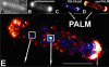
The Escherichia coli chemotaxis network is a model system for biological signal processing. In E. coli, transmembrane receptors responsible for signal transduction assemble into large clusters containing several thousand proteins. These sensory clusters have been observed at cell poles and future division sites. Despite extensive study, it remains unclear how chemotaxis clusters form, what controls cluster size and density, and how the cellular location of clusters is robustly maintained in growing and dividing cells. Here, we use photoactivated localization microscopy (PALM) to map the cellular locations of three proteins central to bacterial chemotaxis (the Tar receptor, CheY, and CheW) with a precision of 15 nm. We find that cluster sizes are approximately exponentially distributed, with no characteristic cluster size. One-third of Tar receptors are part of smaller lateral clusters and not of the large polar clusters. Analysis of the relative cellular locations of 1.1 million individual proteins (from 326 cells) suggests that clusters form via stochastic self-assembly. The super-resolution PALM maps of E. coli receptors support the notion that stochastic self-assembly can create and maintain approximately periodic structures in biological membranes, without direct cytoskeletal involvement or active transport.
Commentary: Our goal as tool developers is to invent methods capable of uncovering new biological insights unobtainable by pre-existing technologies. A terrific example is given by this paper, where grad students Derek Greenfield and Ann McEvoy in Jan Liphardt’s group at Berkeley used our PALM to image the size and position distributions of chemotaxis proteins in E. Coli with unprecedented precision and sensitivity. Their analysis revealed that the cluster sizes follow a stretched exponential distribution, and the density of clusters is highest furthest away from the largest (e.g., polar) clusters. Both observations support a model for passive self-assembly rather than active cytoskeletal assembly of the chemotaxis network.
The pH-dependent binding of Igs to the neonatal FcR (FcRn) plays a critical role in the in vivo homeostasis of IgGs. Modulating the interaction between Fc and FcRn through protein engineering is one method for improving the pharmacokinetics of therapeutic Abs. Recent studies disputed the direct relationship between increasing FcRn affinity and improved pharmacokinetic properties. In this work, we studied the pharmacokinetics of two human IgG1 Fc variants in cynomolgus monkey to further clarify the affinity-pharmacokinetic relationship. First, we report a number of novel Fc point mutations and combination variants, including some with primate-specific FcRn-binding improvements. By studying these variants along with some previously described variants across a wide range of affinities, we discovered a direct correlation of pH 6 affinity improvements with neutral pH improvements, suggesting that all of the tested variants exhibit similar pH dependency in FcRn binding. We then evaluated the pharmacokinetics of variants N434A and N434W, which, respectively, gave approximately 4- and 80-fold improvements in pH 6-binding affinity to both human and nonhuman primate FcRn. Surprisingly, clearance of N434W was similar to that of wild type. N434W is the first variant studied in primates that exhibits significant binding to FcRn at pH 7.4, and its clearance substantiates the principle that too much affinity improvement, i.e., beyond that of N434W, does not yield improved pharmacokinetics. In contrast, N434A exhibited a approximately 2-fold decrease in clearance in cynomolgus monkey, supporting the notion that modest increases in pH 6 FcRn affinity can result in improved pharmacokinetics in primates.
The central actions of leptin are essential for homeostatic control of adipose tissue mass, glucose metabolism, and many autonomic and neuroendocrine systems. In the brain, leptin acts on numerous different cell types via the long-form leptin receptor (LepRb) to elicit its effects. The precise identification of leptin’s cellular targets is fundamental to understanding the mechanism of its pleiotropic central actions. We have systematically characterized LepRb distribution in the mouse brain using in situ hybridization in wildtype mice as well as by EYFP immunoreactivity in a novel LepRb-IRES-Cre EYFP reporter mouse line showing high levels of LepRb mRNA/EYFP coexpression. We found substantial LepRb mRNA and EYFP expression in hypothalamic and extrahypothalamic sites described before, including the dorsomedial nucleus of the hypothalamus, ventral premammillary nucleus, ventral tegmental area, parabrachial nucleus, and the dorsal vagal complex. Expression in insular cortex, lateral septal nucleus, medial preoptic area, rostral linear nucleus, and in the Edinger-Westphal nucleus was also observed and had been previously unreported. The LepRb-IRES-Cre reporter line was used to chemically characterize a population of leptin receptor-expressing neurons in the midbrain. Tyrosine hydroxylase and Cre reporter were found to be coexpressed in the ventral tegmental area and in other midbrain dopaminergic neurons. Lastly, the LepRb-IRES-Cre reporter line was used to map the extent of peripheral leptin sensing by central nervous system (CNS) LepRb neurons. Thus, we provide data supporting the use of the LepRb-IRES-Cre line for the assessment of the anatomic and functional characteristics of neurons expressing leptin receptor.
We have demonstrated super-resolution imaging of protein distributions in cells at depth at multiple layers with a lateral localization precision better than 50 nm. The approach is based on combining photoactivated localization microscopy with temporal focusing.
To grasp the international developing tendency of acupuncture research and provide some references for promoting acupuncture and moxibustion internationalization process, the articles about acupuncture in Science Citation Index (SCI) periodicals in 2007 were retrieved by adopting the retrieval tactics on line in combination with database searching. Results indicate that 257 articles about acupuncture had been retrived from the SCI Web databases. These articles were published in 125 journals respectively, most of which were Euramerican journals. Among these journals, the impact factor of the Journal of the American Medical Association (JAMA), 25. 547, is the highest one. It is shown that the impact factors of the SCI periodicals, in which acupuncture articles embodied are increased, the quality of these articles are improved obviously and the types of the articles are various in 2007, but there is obvious difference in the results of these studies due to the difference of experimental methods, the subjects of these experiments and acupuncture manipulations. Therefore, standardization of many problems arising from the researches on acupuncture is extremely imminent.
Gene expression depends upon the antagonistic actions of chromatin remodeling complexes. While this has been studied extensively for the enzymes that covalently modify the tails of histones, the mechanism of how ATP-dependent remodeling complexes antagonize each other to maintain the proper level of gene activity is not known. The gene encoding a large subunit of ribonucleotide reductase, RNR3, is regulated by ISW2 and SWI/SNF, complexes that repress and activate transcription, respectively. Here, we studied the functional interactions of these two complexes at RNR3. Deletion of ISW2 causes constitutive recruitment of SWI/SNF, and conditional reexpression of ISW2 causes the repositioning of nucleosomes and reduced SWI/SNF occupancy at RNR3. Thus, ISW2 is required for restriction of access of SWI/SNF to the RNR3 promoter under the uninduced condition. Interestingly, the binding of sequence-specific DNA binding factors and the general transcription machinery are unaffected by the status of ISW2, suggesting that disruption of nucleosome positioning does not cause a nonspecific increase in cross-linking of all factors to RNR3. We provide evidence that ISW2 does not act on SWI/SNF directly but excludes its occupancy by positioning nucleosomes over the promoter. Genetic disruption of nucleosome positioning by other means led to a similar phenotype, linking repressed chromatin structure to SWI/SNF exclusion. Thus, incorporation of promoters into a repressive chromatin structure is essential for prevention of the opportunistic actions of nucleosome-disrupting activities in vivo, providing a novel mechanism for maintaining tight control of gene expression.