Filter
Associated Lab
- Ahrens Lab (1) Apply Ahrens Lab filter
- Dickson Lab (1) Apply Dickson Lab filter
- Gonen Lab (1) Apply Gonen Lab filter
- Heberlein Lab (1) Apply Heberlein Lab filter
- Keller Lab (1) Apply Keller Lab filter
- Lippincott-Schwartz Lab (1) Apply Lippincott-Schwartz Lab filter
- Satou Lab (1) Apply Satou Lab filter
- Singer Lab (1) Apply Singer Lab filter
Publication Date
- May 30, 2013 (1) Apply May 30, 2013 filter
- May 23, 2013 (1) Apply May 23, 2013 filter
- May 20, 2013 (1) Apply May 20, 2013 filter
- May 13, 2013 (1) Apply May 13, 2013 filter
- May 12, 2013 (1) Apply May 12, 2013 filter
- May 9, 2013 (1) Apply May 9, 2013 filter
- May 8, 2013 (1) Apply May 8, 2013 filter
- May 1, 2013 (3) Apply May 1, 2013 filter
- Remove May 2013 filter May 2013
- Remove 2013 filter 2013
Type of Publication
10 Publications
Showing 1-10 of 10 resultsHow the brain perceives sensory information and generates meaningful behavior depends critically on its underlying circuitry. The protocerebral bridge (PB) is a major part of the insect central complex (CX), a premotor center that may be analogous to the human basal ganglia. Here, by deconstructing hundreds of PB single neurons and reconstructing them into a common three-dimensional framework, we have constructed a comprehensive map of PB circuits with labeled polarity and predicted directions of information flow. Our analysis reveals a highly ordered information processing system that involves directed information flow among CX subunits through 194 distinct PB neuron types. Circuitry properties such as mirroring, convergence, divergence, tiling, reverberation, and parallel signal propagation were observed; their functional and evolutional significance is discussed. This layout of PB neuronal circuitry may provide guidelines for further investigations on transformation of sensory (e.g., visual) input into locomotor commands in fly brains.
Many species are critically dependent on olfaction for survival. In the main olfactory system of mammals, odours are detected by sensory neurons that express a large repertoire of canonical odorant receptors and a much smaller repertoire of trace amine-associated receptors (TAARs). Odours are encoded in a combinatorial fashion across glomeruli in the main olfactory bulb, with each glomerulus corresponding to a specific receptor. The degree to which individual receptor genes contribute to odour perception is unclear. Here we show that genetic deletion of the olfactory Taar gene family, or even a single Taar gene (Taar4), eliminates the aversion that mice display to low concentrations of volatile amines and to the odour of predator urine. Our findings identify a role for the TAARs in olfaction, namely, in the high-sensitivity detection of innately aversive odours. In addition, our data reveal that aversive amines are represented in a non-redundant fashion, and that individual main olfactory receptor genes can contribute substantially to odour perception.
During locomotion in vertebrates, reticulospinal neurons in the hindbrain play critical roles in providing descending excitation to the spinal cord locomotor systems. However, despite the fact that many genes that are used to classify the neuronal identities of neurons in the hindbrain have been identified, the molecular identity of the reticulospinal neurons that are critically involved in locomotor drive is not well understood. Chx10-expressing neurons (V2a neurons) are ipsilaterally projecting glutamatergic neurons in the spinal cord and the hindbrain. Many of the V2a neurons in the hindbrain are known to project to the spinal cord in zebrafish, making hindbrain V2a neurons a prime candidate in descending locomotor drive. Results We investigated the roles of hindbrain V2a neurons using optogenetic and electrophysiological approaches. The forced activation of hindbrain V2a neurons using channelrhodopsin efficiently evoked swimming, whereas the forced inactivation of them using Archearhodopsin3 or Halorhodpsin reliably stopped ongoing swimming. Electrophysiological recordings of two populations of hindbrain reticulospinal V2a neurons showed that they were active during swimming. One population of neurons, small V2a neurons in the caudal hindbrain, fired with low rhythmicity, whereas the other population of neurons, large reticulospinal V2a neurons, called MiV1 neurons, fired more rhythmically. Conclusions These results indicated that hindbrain reticulospinal V2a neurons play critical roles in providing excitation to the spinal locomotor circuits during swimming by providing both tonic and phasic inputs to the circuits.
In rats, navigating through an environment requires continuous information about objects near the head. Sensory information such as object location and surface texture are encoded by spike firing patterns of single neurons within rat barrel cortex. Although there are many studies using single-unit electrophysiology, much less is known regarding the spatiotemporal pattern of activity of populations of neurons in barrel cortex in response to whisker stimulation. To examine cortical response at the population level, we used voltage-sensitive dye (VSD) imaging to examine ensemble spatiotemporal dynamics of barrel cortex in response to stimulation of single or two adjacent whiskers in urethane-anesthetized rats. Single whisker stimulation produced a poststimulus fluorescence response peak within 12-16 ms in the barrel corresponding to the stimulated whisker (principal whisker). This fluorescence subsequently propagated throughout the barrel field, spreading anisotropically preferentially along a barrel row. After paired whisker stimulation, the VSD signal showed sublinear summation (less than the sum of 2 single whisker stimulations), consistent with previous electrophysiological and imaging studies. Surprisingly, we observed a spatial shift in the center of activation occurring over a 10- to 20-ms period with shift magnitudes of 1-2 barrels. This shift occurred predominantly in the posteromedial direction within the barrel field. Our data thus reveal previously unreported spatiotemporal patterns of barrel cortex activation. We suggest that this nontopographical shift is consistent with known functional and anatomic asymmetries in barrel cortex and that it may provide an important insight for understanding barrel field activation during whisking behavior.
Mineral nitrogen in nature is often found in the form of nitrate (NO3(-)). Numerous microorganisms evolved to assimilate nitrate and use it as a major source of mineral nitrogen uptake. Nitrate, which is central in nitrogen metabolism, is first reduced to nitrite (NO2(-)) through a two-electron reduction reaction. The accumulation of cellular nitrite can be harmful because nitrite can be reduced to the cytotoxic nitric oxide. Instead, nitrite is rapidly removed from the cell by channels and transporters, or reduced to ammonium or dinitrogen through the action of assimilatory enzymes. Despite decades of effort no structure is currently available for any nitrate transport protein and the mechanism by which nitrate is transported remains largely unknown. Here we report the structure of a bacterial nitrate/nitrite transport protein, NarK, from Escherichia coli, with and without substrate. The structures reveal a positively charged substrate-translocation pathway lacking protonatable residues, suggesting that NarK functions as a nitrate/nitrite exchanger and that protons are unlikely to be co-transported. Conserved arginine residues comprise the substrate-binding pocket, which is formed by association of helices from the two halves of NarK. Key residues that are important for substrate recognition and transport are identified and related to extensive mutagenesis and functional studies. We propose that NarK exchanges nitrate for nitrite by a rocker switch mechanism facilitated by inter-domain hydrogen bond networks.
The Influenza A virus genome consists of eight negative sense, single-stranded RNA segments. Although it has been established that most virus particles contain a single copy of each of the eight viral RNAs, the packaging selection mechanism remains poorly understood. Influenza viral RNAs are synthesized in the nucleus, exported into the cytoplasm and travel to the plasma membrane where viral budding and genome packaging occurs. Due to the difficulties in analyzing associated vRNPs while preserving information about their positions within the cell, it has remained unclear how and where during cellular trafficking the viral RNAs of different segments encounter each other. Using a multicolor single-molecule sensitivity fluorescence in situ hybridization (smFISH) approach, we have quantitatively monitored the colocalization of pairs of influenza viral RNAs in infected cells. We found that upon infection, the viral RNAs from the incoming particles travel together until they reach the nucleus. The viral RNAs were then detected in distinct locations in the nucleus; they are then exported individually and initially remain separated in the cytoplasm. At later time points, the different viral RNA segments gather together in the cytoplasm in a microtubule independent manner. Viral RNAs of different identities colocalize at a high frequency when they are associated with Rab11 positive vesicles, suggesting that Rab11 positive organelles may facilitate the association of different viral RNAs. Using engineered influenza viruses lacking the expression of HA or M2 protein, we showed that these viral proteins are not essential for the colocalization of two different viral RNAs in the cytoplasm. In sum, our smFISH results reveal that the viral RNAs travel together in the cytoplasm before their arrival at the plasma membrane budding sites. This newly characterized step of the genome packaging process demonstrates the precise spatiotemporal regulation of the infection cycle.
In both mammalian and insect models of ethanol intoxication, high doses of ethanol induce motor impairment and eventually sedation. Sensitivity to the sedative effects of ethanol is inversely correlated with risk for alcoholism. However, the genes regulating ethanol sensitivity are largely unknown. Based on a previous genetic screen in Drosophila for ethanol sedation mutants, we identified a novel gene, tank (CG15626), the homolog of the mammalian tumor suppressor EI24/PIG8, which has a strong role in regulating ethanol sedation sensitivity. Genetic and behavioral analyses revealed that tank acts in the adult nervous system to promote ethanol sensitivity. We localized the function of tank in regulating ethanol sensitivity to neurons within the pars intercerebralis that have not been implicated previously in ethanol responses. We show that acutely manipulating the activity of all tank-expressing neurons, or of pars intercerebralis neurons in particular, alters ethanol sensitivity in a sexually dimorphic manner, since neuronal activation enhanced ethanol sedation in males, but not females. Finally, we provide anatomical evidence that tank-expressing neurons form likely synaptic connections with neurons expressing the neural sex determination factor fruitless (fru), which have been implicated recently in the regulation of ethanol sensitivity. We suggest that a functional interaction with fru neurons, many of which are sexually dimorphic, may account for the sex-specific effect induced by activating tank neurons. Overall, we have characterized a novel gene and corresponding set of neurons that regulate ethanol sensitivity in Drosophila.
The glucose transporter, GLUT4, redistributes to the plasma membrane (PM) upon insulin stimulation, but also recycles through endosomal compartments. Different Rab proteins control these transport itineraries of GLUT4. However, the specific roles played by different Rab proteins in GLUT4 trafficking has been difficult to assess, primarily due to the complexity of endomembrane organization and trafficking. To address this problem, we recently performed advanced live cell imaging using total internal reflection fluorescence (TIRF) microscopy, which images objects ~150 nm from the PM, directly visualizing GLUT4 trafficking in response to insulin stimulation. Using IRAP-pHluorin to selectively label GSVs undergoing PM fusion in response to insulin, we identified Rab10 as the only Rab protein that binds this compartment. Rab14 was found to label transferrin-positive, endosomal compartments containing GLUT4. These also could fuse with the PM in response to insulin, albeit more slowly. Several other Rab proteins, including Rab4A, 4B and 8A, were found to mediate GLUT4 intra-endosomal recycling, serving to internalize surface-bound GLUT4 into endosomal compartments for ultimate delivery to GSVs. Thus, multiple Rab proteins regulate the circulation of GLUT4 molecules within the endomembrane system, maintaining optimal insulin responsiveness within cells.
Transcription factors that can convert adult cells of one type to another are usually discovered empirically by testing factors with a known developmental role in the target cell. Here we show that standard genomic methods (RNA-seq and ChIP-seq) can help identify these factors, as most are more strongly Polycomb repressed in the source cell and more highly expressed in the target cell. This criterion is an effective genome-wide screen that significantly enriches for factors that can transdifferentiate several mammalian cell types including neural stem cells, neurons, pancreatic islets, and hepatocytes. These results suggest that barriers between adult cell types, as depicted in Waddington’s "epigenetic landscape", consist in part of differentially Polycomb-repressed transcription factors. This genomic model of cell identity helps rationalize a growing number of transdifferentiation protocols and may help facilitate the engineering of cell identity for regenerative medicine.
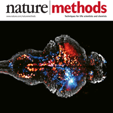
Brain function relies on communication between large populations of neurons across multiple brain areas, a full understanding of which would require knowledge of the time-varying activity of all neurons in the central nervous system. Here we use light-sheet microscopy to record activity, reported through the genetically encoded calcium indicator GCaMP5G, from the entire volume of the brain of the larval zebrafish in vivo at 0.8 Hz, capturing more than 80% of all neurons at single-cell resolution. Demonstrating how this technique can be used to reveal functionally defined circuits across the brain, we identify two populations of neurons with correlated activity patterns. One circuit consists of hindbrain neurons functionally coupled to spinal cord neuropil. The other consists of an anatomically symmetric population in the anterior hindbrain, with activity in the left and right halves oscillating in antiphase, on a timescale of 20 s, and coupled to equally slow oscillations in the inferior olive.

