The volume imaged is given by: Volume = VoxelSize x VoxelRate x Duration. The voxel size is set as just described to (8-10 nm)3 . The voxel rate is likewise empirically determined by the reconstruction difficulty for different signal to noise ratios (SNRs). In our case of fly tissue we have found that a minimum signal to noise ratio of ~6 is required. This SNR is in turn determined by the primary beam current, sample staining strength, and the physics of the electron scattering and their detection. We have just achieved a 3x improvement in acquisition rate by switching to the Zeiss Merlin SEM which can sustain a primary electron current of close to 8 nanoamps, versus 2 nanoamps of the standard SEM, with only tolerable compromise of x,y beam resolution.
The most significant improvements have been made in the last item of the expression, the duration of seamless acquisition. Initially we could acquire data for only a few days before an uncontrolled termination event, usually a FIB column failure. Now we routinely run for 2-3 months and only stop when the sample imaging has been completed. This required addressing a variety of interrupt issues: ion source reheat, utility failure (water, power, air, and temperature fluctuation), and microscope failure (focus, electrical, software, vacuum). With improvements and backups for the existing utilities, and with the transition to a new lab space with special environmental and power backup, we have decreased the frequency of these problems. Close monitoring of major FIB-SEM parameters - beam current, focus, and so on - enables us to shut down safely in the case of many remaining failure events. Finally a feedback scheme that controls the milling ion beam enables us to seamlessly restart without losing milling control that otherwise would result in a loss of 100 nm of sample thickness. The seamless restarting capability effectively removes any fixed volume limit due to interruptions.
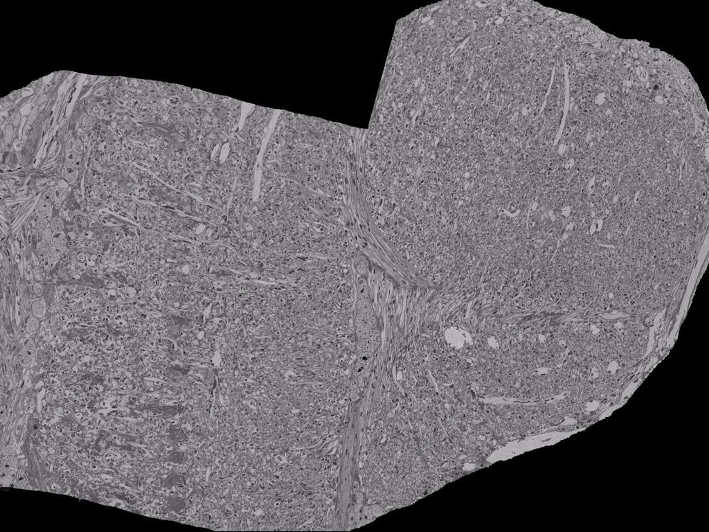
Figure 1: Medulla-Lobula-Lobular Plate sample
As an example of improved capabilities, the FIB-SEM data set shown in Fig. 1, covers a major cross section of the medulla and full cross section of the lobula and lobula plate. It was taken over a 3 month period and illustrates the size and quality of neuropile that can now be imaged. Several similar sized volumes of complete antennal lobes have also been imaged on a routine basis. Such images are typically taken of several samples to find the best possible stain, contrast, synapse clarity, and membrane integrity to minimize the large proof-reading time investment. Three FIB-SEM machines, the original Zeiss NVision and two FEI/Merlin (with 2-3 x throughput) are now in production mode for various biological targets.