Filter
Associated Lab
Associated Project Team
6 Janelia Publications
Showing 1-6 of 6 resultsHigh-resolution microscopic imaging of biological samples often produces multiple 3D image tiles to cover a large field of view of specimen. Usually each tile has a large size, in the range of hundreds of megabytes to several gigabytes. For many of our image data sets, existing software tools are often unable to stitch those 3D tiles into a panoramic view, thus impede further data analysis. We propose a simple, but accurate, robust, and automatic method to stitch a group of image tiles without a priori adjacency information of them. We first use a multiscale strategy to register a pair of 3D image tiles rapidly, achieving about 8~10 times faster speed and 10 times less memory requirement compared to previous methods. Then we design a minimum-spanning-tree based method to determine the optimal adjacency of tiles. We have successfully stitched large image stacks of model animals including C. elegans, fruit fly, dragonfly, and mouse, which could not be stitched by several existing methods.
A parallel wavefront optimization method is demonstrated experimentally to focus light through random scattering media. The simultaneous modulation of multiple phase elements, each at a unique frequency, enables a parallel determination of the optimal wavefront. Compared to a pixel-by-pixel measurement, the reported parallel method uses the target signal in a highly efficient way. With 441 phase elements, a high-quality focus was formed through a glass diffuser with a peak-to-background ratio of \~{}270. The accuracy and repeatability of the system were tested through experiments.

Recent findings implicate alternate core promoter recognition complexes in regulating cellular differentiation. Here we report a spatial segregation of the alternative core factor TAF3, but not canonical TFIID subunits, away from the nuclear periphery, where the key myogenic gene MyoD is preferentially localized in myoblasts. This segregation is correlated with the differential occupancy of TAF3 versus TFIID at the MyoD promoter. Loss of this segregation by modulating either the intranuclear location of the MyoD gene or TAF3 protein leads to altered TAF3 occupancy at the MyoD promoter. Intriguingly, in differentiated myotubes, the MyoD gene is repositioned to the nuclear interior, where TAF3 resides. The specific high-affinity recognition of H3K4Me3 by the TAF3 PHD (plant homeodomain) finger appears to be required for the sequestration of TAF3 to the nuclear interior. We suggest that intranuclear sequestration of core transcription components and their target genes provides an additional mechanism for promoter selectivity during differentiation.
Commentary: Jie Yao in Bob Tijan’s lab used a combination of confocal microscopy and dual label PALM in thin sections cut from resin-embedded cells to show that certain core transcription components and their target genes are spatially segregated in myoblasts, but not in differentiated myotubes, suggesting that such spatial segregation may play a role in guiding cellular differentiation.
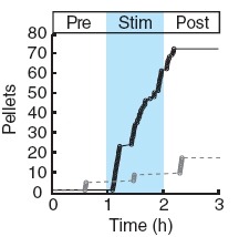
Two intermingled hypothalamic neuron populations specified by expression of agouti-related peptide (AGRP) or pro-opiomelanocortin (POMC) positively and negatively influence feeding behavior, respectively, possibly by reciprocally regulating downstream melanocortin receptors. However, the sufficiency of these neurons to control behavior and the relationship of their activity to the magnitude and dynamics of feeding are unknown. To measure this, we used channelrhodopsin-2 for cell type-specific photostimulation. Activation of only 800 AGRP neurons in mice evoked voracious feeding within minutes. The behavioral response increased with photoexcitable neuron number, photostimulation frequency and stimulus duration. Conversely, POMC neuron stimulation reduced food intake and body weight, which required melanocortin receptor signaling. However, AGRP neuron-mediated feeding was not dependent on suppressing this melanocortin pathway, indicating that AGRP neurons directly engage feeding circuits. Furthermore, feeding was evoked selectively over drinking without training or prior photostimulus exposure, which suggests that AGRP neurons serve a dedicated role coordinating this complex behavior.
We developed a multicolor neuron labeling technique in Drosophila melanogaster that combines the power to specifically target different neural populations with the label diversity provided by stochastic color choice. This adaptation of vertebrate Brainbow uses recombination to select one of three epitope-tagged proteins detectable by immunofluorescence. Two copies of this construct yield six bright, separable colors. We used Drosophila Brainbow to study the innervation patterns of multiple antennal lobe projection neuron lineages in the same preparation and to observe the relative trajectories of individual aminergic neurons. Nerve bundles, and even individual neurites hundreds of micrometers long, can be followed with definitive color labeling. We traced motor neurons in the subesophageal ganglion and correlated them to neuromuscular junctions to identify their specific proboscis muscle targets. The ability to independently visualize multiple lineage or neuron projections in the same preparation greatly advances the goal of mapping how neurons connect into circuits.
Most neurons of the central complex belong to 10 secondary (larvally produced) lineages. In the late larva, undifferentiated axon tracts of these lineages form a primordium in which all of the compartments of the central complex can be recognized as discrete entities. Four posterior lineages (DPMm1, DPMpm1, DPMpm2, and CM4) generate the classes of small-field neurons that interconnect the protocerebral bridge, fan-shaped body, noduli, and ellipsoid body. Three lineages located in the anterior brain, DALv2, BAmv1, and DALcl2, form the large-field neurons of the ellipsoid body and fan-shaped body, respectively. These lineages provide an input channel from the optic tubercle and connect the central complex with adjacent anterior brain compartments. Three lineages in the posterior cortex, CM3, CP2, and DPMpl2, connect the posterior brain neuropil with specific layers of the fan-shaped body. Even though all of the compartments of the central complex are prefigured in the late larval brain by the axon tracts of the above-mentioned lineages, the neuropil differentiates during the first 2 days of the pupal period when terminal branches and synapses of secondary neurons are formed. During this phase the initially straight horizontal layers of the central complex bend in the frontal plane, which produces the characteristic shape of the fan-shaped and ellipsoid body. Our analysis provides a comprehensive picture of the lineages that form the central complex, and will facilitate future studies that address the structure or function of the central complex at the single cell level.