Filter
Associated Lab
- Aso Lab (1) Apply Aso Lab filter
- Baker Lab (3) Apply Baker Lab filter
- Betzig Lab (7) Apply Betzig Lab filter
- Bock Lab (1) Apply Bock Lab filter
- Cui Lab (3) Apply Cui Lab filter
- Dickson Lab (1) Apply Dickson Lab filter
- Dudman Lab (1) Apply Dudman Lab filter
- Eddy/Rivas Lab (5) Apply Eddy/Rivas Lab filter
- Fetter Lab (2) Apply Fetter Lab filter
- Gonen Lab (1) Apply Gonen Lab filter
- Hess Lab (2) Apply Hess Lab filter
- Jayaraman Lab (2) Apply Jayaraman Lab filter
- Ji Lab (2) Apply Ji Lab filter
- Keller Lab (2) Apply Keller Lab filter
- Lavis Lab (5) Apply Lavis Lab filter
- Lee (Albert) Lab (1) Apply Lee (Albert) Lab filter
- Leonardo Lab (2) Apply Leonardo Lab filter
- Looger Lab (7) Apply Looger Lab filter
- Magee Lab (1) Apply Magee Lab filter
- Menon Lab (3) Apply Menon Lab filter
- Murphy Lab (1) Apply Murphy Lab filter
- Reiser Lab (2) Apply Reiser Lab filter
- Riddiford Lab (1) Apply Riddiford Lab filter
- Rubin Lab (3) Apply Rubin Lab filter
- Scheffer Lab (2) Apply Scheffer Lab filter
- Schreiter Lab (2) Apply Schreiter Lab filter
- Simpson Lab (3) Apply Simpson Lab filter
- Singer Lab (1) Apply Singer Lab filter
- Sternson Lab (6) Apply Sternson Lab filter
- Svoboda Lab (7) Apply Svoboda Lab filter
- Tjian Lab (2) Apply Tjian Lab filter
- Truman Lab (1) Apply Truman Lab filter
- Zlatic Lab (1) Apply Zlatic Lab filter
- Zuker Lab (2) Apply Zuker Lab filter
Associated Project Team
Publication Date
- December 2011 (10) Apply December 2011 filter
- November 2011 (8) Apply November 2011 filter
- October 2011 (8) Apply October 2011 filter
- September 2011 (8) Apply September 2011 filter
- August 2011 (9) Apply August 2011 filter
- July 2011 (5) Apply July 2011 filter
- June 2011 (10) Apply June 2011 filter
- May 2011 (6) Apply May 2011 filter
- April 2011 (5) Apply April 2011 filter
- March 2011 (6) Apply March 2011 filter
- February 2011 (8) Apply February 2011 filter
- January 2011 (15) Apply January 2011 filter
- Remove 2011 filter 2011
98 Janelia Publications
Showing 31-40 of 98 resultsWe took advantage of the unusual genomic organization of the ciliate Oxytricha trifallax to screen for eukaryotic non-coding RNA (ncRNA) genes. Ciliates have two types of nuclei: a germ line micronucleus that is usually transcriptionally inactive, and a somatic macronucleus that contains a reduced, fragmented and rearranged genome that expresses all genes required for growth and asexual reproduction. In some ciliates including Oxytricha, the macronuclear genome is particularly extreme, consisting of thousands of tiny ’nanochromosomes’, each of which usually contains only a single gene. Because the organism itself identifies and isolates most of its genes on single-gene nanochromosomes, nanochromosome structure could facilitate the discovery of unusual genes or gene classes, such as ncRNA genes. Using a draft Oxytricha genome assembly and a custom-written protein-coding genefinding program, we identified a subset of nanochromosomes that lack any detectable protein-coding gene, thereby strongly enriching for nanochromosomes that carry ncRNA genes. We found only a small proportion of non-coding nanochromosomes, suggesting that Oxytricha has few independent ncRNA genes besides homologs of already known RNAs. Other than new members of known ncRNA classes including C/D and H/ACA snoRNAs, our screen identified one new family of small RNA genes, named the Arisong RNAs, which share some of the features of small nuclear RNAs.
Because of its genetic, molecular, and behavioral tractability, Drosophila has emerged as a powerful model system for studying molecular and cellular mechanisms underlying the development and function of nervous systems. The Drosophila nervous system has fewer neurons and exhibits a lower glia:neuron ratio than is seen in vertebrate nervous systems. Despite the simplicity of the Drosophila nervous system, glial organization in flies is as sophisticated as it is in vertebrates. Furthermore, fly glial cells play vital roles in neural development and behavior. In addition, powerful genetic tools are continuously being created to explore cell function in vivo. In taking advantage of these features, the fly nervous system serves as an excellent model system to study general aspects of glial cell development and function in vivo. In this article, we review and discuss advanced genetic tools that are potentially useful for understanding glial cell biology in Drosophila.
Parallel circuits throughout the CNS exhibit distinct sensitivities and responses to sensory stimuli. Ambiguities in the source and properties of signals elicited by physiological stimuli, however, frequently obscure the mechanisms underlying these distinctions. We found that differences in the degree to which activity in two classes of Off retinal ganglion cell (RGC) encode information about light stimuli near detection threshold were not due to obvious differences in the cells’ intrinsic properties or the chemical synaptic input the cells received; indeed, differences in the cells’ light responses were largely insensitive to block of fast ionotropic glutamate receptors. Instead, the distinct responses of the two types of RGCs likely reflect differences in light-evoked electrical synaptic input. These results highlight a surprising strategy by which the retina differentially processes and routes visual information and provide new insight into the circuits that underlie responses to stimuli near detection threshold.
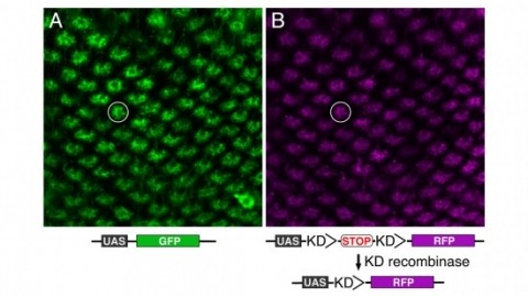
Site-specific recombinases have been used for two decades to manipulate the structure of animal genomes in highly predictable ways and have become major research tools. However, the small number of recombinases demonstrated to have distinct specificities, low toxicity, and sufficient activity to drive reactions to completion in animals has been a limitation. In this report we show that four recombinases derived from yeast-KD, B2, B3, and R-are highly active and nontoxic in Drosophila and that KD, B2, B3, and the widely used FLP recombinase have distinct target specificities. We also show that the KD and B3 recombinases are active in mice.
MOTIVATION: Automatic recognition of cell identities is critical for quantitative measurement, targeting, and manipulation of cells of model animals at single-cell resolution. It has been shown to be a powerful tool for studying gene expression and regulation, cell lineages, and cell fates. Existing methods first segment cells, before applying a recognition algorithm in the second step. As a result, the segmentation errors in the first step directly affect and complicate the subsequent cell recognition step. Moreover, in new experimental settings, some of the image features that have been previously relied upon to recognize cells may not be easy to reproduce, due to limitations on the number of color channels available for fluorescent imaging or to the cost of building transgenic animals. An approach that is more accurate and relies on only a single signal channel is clearly desirable. RESULTS: We have developed a new method, called SRS (for Simultaneous Recognition and Segmentation of cells), and applied it to 3D image stacks of the model organism C. elegans. Given a 3D image stack of the animal and a 3D atlas of target cells, SRS is effectively an atlas-guided voxel classification process: cell recognition is realized by smoothly deforming the atlas to best fit the image, where the segmentation is obtained naturally via classification of all image voxels. The method achieved a 97.7% overall recognition accuracy in recognizing a key class of marker cells, the body wall muscle (BWM) cells, on a data set of 175 C. elegans image stacks containing 14,118 manually curated BWM cells providing the "ground-truth" for accuracy. This result was achieved without any additional fiducial image features. SRS also automatically identified 14 of the image stacks as involving ±90-degree rotations. With these stacks excluded from the data set, the recognition accuracy rose to 99.1%. We also show SRS is generally applicable to other cell-types, e.g. intestinal cells. AVAILABILITY: The supplementary movies can be downloaded from our website http://penglab.janelia.org/proj/celegans_seganno. The method has been implemented as a plug-in program within the V3D system (http://penglab.janelia.org/proj/v3d) and will be released in the V3D plugin source code repository.
Pavlovian olfactory learning in Drosophila produces two genetically distinct forms of intermediate-term memories: anesthesia-sensitive memory, which requires the amnesiac gene, and anesthesia-resistant memory (ARM), which requires the radish gene. Here, we report that ARM is specifically enhanced or inhibited in flies with elevated or reduced serotonin (5HT) levels, respectively. The requirement for 5HT was additive with the memory defect of the amnesiac mutation but was occluded by the radish mutation. This result suggests that 5HT and Radish protein act on the same pathway for ARM formation. Three supporting lines of evidence indicate that ARM formation requires 5HT released from only two dorsal paired medial (DPM) neurons onto the mushroom bodies (MBs), the olfactory learning and memory center in Drosophila: (i) DPM neurons were 5HT-antibody immunopositive; (ii) temporal inhibition of 5HT synthesis or release from DPM neurons, but not from other serotonergic neurons, impaired ARM formation; (iii) knocking down the expression of d5HT1A serotonin receptors in α/β MB neurons, which are innervated by DPM neurons, inhibited ARM formation. Thus, in addition to the Amnesiac peptide required for anesthesia-sensitive memory formation, the two DPM neurons also release 5HT acting on MB neurons for ARM formation.
The ways in which cells set the size of intracellular structures is an important but largely unsolved problem [1]. Early embryonic divisions pose special problems in this regard. Many checkpoints common in somatic cells are missing from these divisions, which are characterized by rapid reductions in cell size and short cell cycles [2]. Embryonic cells must therefore possess simple and robust mechanisms that allow the size of many of their intracellular structures to rapidly scale with cell size.
Bacterial Rho-independent terminators (RITs) are important genomic landmarks involved in gene regulation and terminating gene expression. In this investigation we present RNIE, a probabilistic approach for predicting RITs. The method is based upon covariance models which have been known for many years to be the most accurate computational tools for predicting homology in structural non-coding RNAs. We show that RNIE has superior performance in model species from a spectrum of bacterial phyla. Further analysis of species where a low number of RITs were predicted revealed a highly conserved structural sequence motif enriched near the genic termini of the pathogenic Actinobacteria, Mycobacterium tuberculosis. This motif, together with classical RITs, account for up to 90% of all the significantly structured regions from the termini of M. tuberculosis genic elements. The software, predictions and alignments described below are available from http://github.com/ppgardne/RNIE.