Filter
Associated Lab
4 Janelia Publications
Showing 1-4 of 4 resultsThe mammalian vomeronasal organ encodes pheromone information about gender, reproductive status, genetic background and individual differences. It remains unknown how pheromone information interacts to trigger innate behaviors. In this study, we identify vomeronasal receptors responsible for detecting female pheromones. A sub-group of V1re clade members recognizes gender-identifying cues in female urine. Multiple members of the V1rj clade are cognate receptors for urinary estrus signals, as well as for sulfated estrogen (SE) compounds. In both cases, the same cue activates multiple homologous receptors, suggesting redundancy in encoding female pheromone cues. Neither gender-specific cues nor SEs alone are sufficient to promote courtship behavior in male mice, whereas robust courtship behavior can be induced when the two cues are applied together. Thus, integrated action of different female cues is required in pheromone-triggered mating behavior. These results suggest a gating mechanism in the vomeronasal circuit in promoting specific innate behavior.DOI: http://dx.doi.org/10.7554/eLife.03025.001.
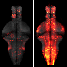
The processing of sensory input and the generation of behavior involves large networks of neurons, which necessitates new technology for recording from many neurons in behaving animals. In the larval zebrafish, light-sheet microscopy can be used to record the activity of almost all neurons in the brain simultaneously at single-cell resolution. Existing implementations, however, cannot be combined with visually driven behavior because the light sheet scans over the eye, interfering with presentation of controlled visual stimuli. Here we describe a system that overcomes the confounding eye stimulation through the use of two light sheets and combines whole-brain light-sheet imaging with virtual reality for fictively behaving larval zebrafish.
Understanding brain function requires monitoring and interpreting the activity of large networks of neurons during behavior. Advances in recording technology are greatly increasing the size and complexity of neural data. Analyzing such data will pose a fundamental bottleneck for neuroscience. We present a library of analytical tools called Thunder built on the open-source Apache Spark platform for large-scale distributed computing. The library implements a variety of univariate and multivariate analyses with a modular, extendable structure well-suited to interactive exploration and analysis development. We demonstrate how these analyses find structure in large-scale neural data, including whole-brain light-sheet imaging data from fictively behaving larval zebrafish, and two-photon imaging data from behaving mouse. The analyses relate neuronal responses to sensory input and behavior, run in minutes or less and can be used on a private cluster or in the cloud. Our open-source framework thus holds promise for turning brain activity mapping efforts into biological insights.
Retinal bipolar cells (BCs) transmit visual signals in parallel channels from the outer to the inner retina, where they provide glutamatergic inputs to specific networks of amacrine and ganglion cells. Intricate network computation at BC axon terminals has been proposed as a mechanism for complex network computation, such as direction selectivity, but direct knowledge of the receptive field property and the synaptic connectivity of the axon terminals of various BC types is required in order to understand the role of axonal computation by BCs. The present study tested the essential assumptions of the presynaptic model of direction selectivity at axon terminals of three functionally distinct BC types that ramify in the direction-selective strata of the mouse retina. Results from two-photon Ca2+ imaging, optogenetic stimulation, and dual patch-clamp recording demonstrated that (1) CB5 cells do not receive fast GABAergic synaptic feedback from starburst amacrine cells (SACs), (2) light-evoked and spontaneous Ca2+ responses are well coordinated among various local regions of CB5 axon terminals, (3) CB5 axon terminals are not directionally selective, (4) CB5 cells consist of two novel functional subtypes with distinct receptive field structures, (5) CB7 cells provide direct excitatory synaptic inputs to, but receive no direct GABAergic synaptic feedback from SACs, and (6) CB7 axon terminals are not directionally selective either. These findings help to simplify models of direction selectivity by ruling out complex computation at BC terminals. They also show that CB5 comprises two functional subclasses of BCs.