Main Menu (Mobile)- Block
Main Menu - Block
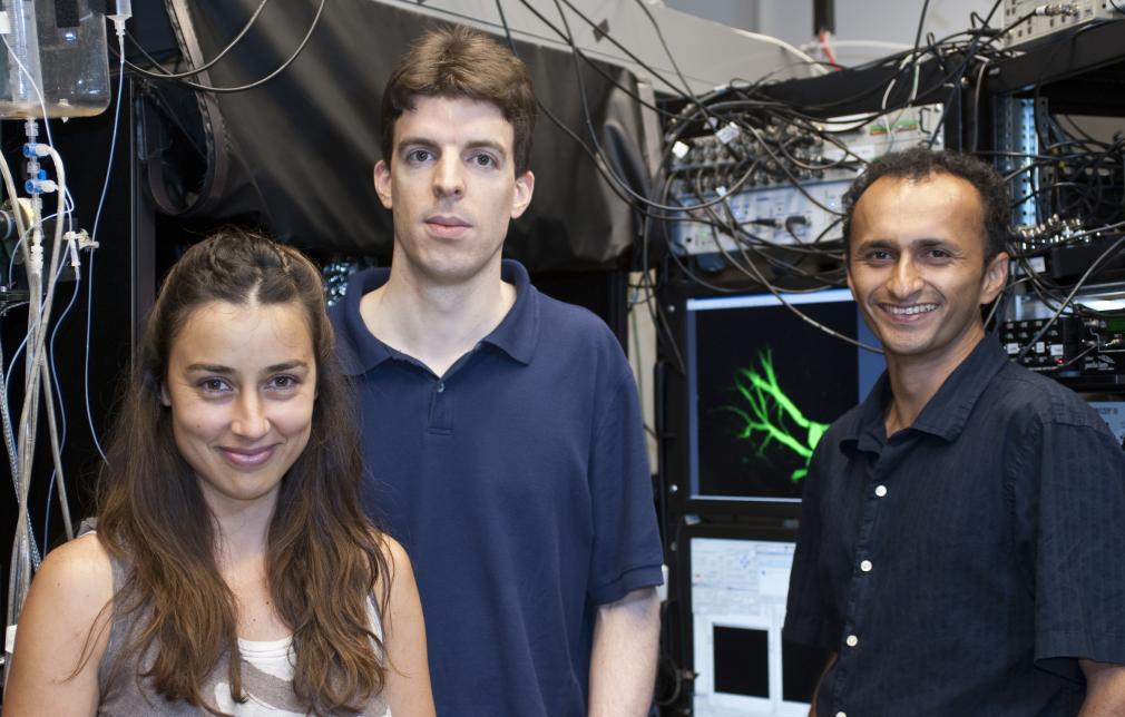
Decision-making during navigation requires processing and integrating multi-sensory information in the context of past experience, internal state and ongoing behavior. Our lab studies such neural dynamics in the fruit fly, Drosophila melanogaster, a genetic model organism that has a small brain but is capable of complex behavior.
Our lab is interested in establishing causal links between the dynamics of neural circuits and the behavioral decisions that an animal continuously makes as it navigates a multi-sensory world. We aim to uncover how the relevant neural representations and dynamics arise and what specific role they play in shaping adaptive behavior.
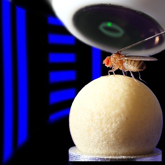
We use the powerful genetic model organism Drosophila melanogaster for our experiments, many of which rely on monitoring and perturbing the activity of specific neural populations during head-fixed behavior. We rely on a combination of two-photon calcium imaging, whole-cell patch clamp electrophysiology, quantitative behavior, optogenetics, and computational analysis and modeling in our efforts to mechanistically link computation in a higher brain region called the central complex to the fly's behavioral decisions.