Main Menu (Mobile)- Block
- Our Research
-
Support Teams
- Overview
- Anatomy and Histology
- Cryo-Electron Microscopy
- Electron Microscopy
- Flow Cytometry
- Fly Facility
- Gene Targeting and Transgenics
- Immortalized Cell Line Culture
- Integrative Imaging
- Janelia Experimental Technology
- Mass Spectrometry
- Media Prep
- Molecular Genomics
- Primary & iPS Cell Culture
- Project Pipeline Support
- Project Technical Resources
- Quantitative Genomics
- Scientific Computing Software
- Scientific Computing Systems
- Viral Tools
- Vivarium
- Open Science
- You + Janelia
- About Us
Main Menu - Block
- Overview
- Anatomy and Histology
- Cryo-Electron Microscopy
- Electron Microscopy
- Flow Cytometry
- Fly Facility
- Gene Targeting and Transgenics
- Immortalized Cell Line Culture
- Integrative Imaging
- Janelia Experimental Technology
- Mass Spectrometry
- Media Prep
- Molecular Genomics
- Primary & iPS Cell Culture
- Project Pipeline Support
- Project Technical Resources
- Quantitative Genomics
- Scientific Computing Software
- Scientific Computing Systems
- Viral Tools
- Vivarium

Demonstrate the power of FIB-SEM data to catalyze biological discoveries.
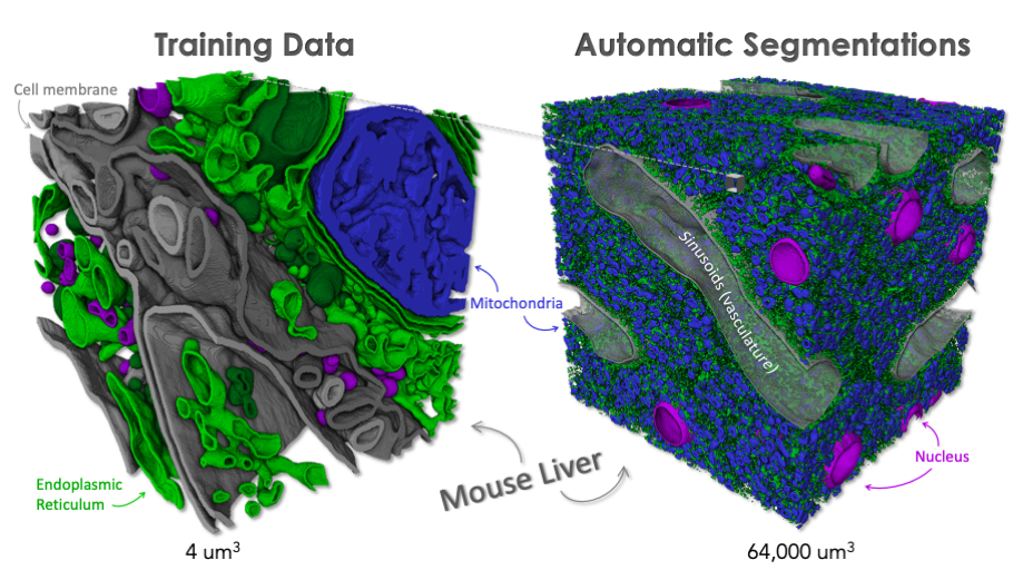
CellMap strives to inspire biological discovery and new insight by showcasing the research potential of 3D architectural and subcellular details at the scale of whole cells and tissues. By generating such 3D data from Focused Ion Beam Scanning Electron Microscopy (FIB-SEM), we can now transcend the traditional 2D cross sectioned electron microscope view of the biological world. This expanded and wholistic 3D view enables new, whole cell characterizations and completely new formulations of hypothesis and questions. CellMap seeks to image samples of broadest general interest across multiple fields in biology, generate initial annotation and segmentation, and share openly so as to catalyze research interest in this new space of 3D biological imaging and data analysis.
To this end, CellMap’s mission is to:
- Produce high-quality, impactful images of diverse cells and tissue samples using FIB-SEMs to study ultrastructure at the cellular level.
- Generate annotations of as many sub-cellular structures as technically possible.
- Make FIB-SEM data, annotations and segmented data, code, sample information, and protocols openly accessible through well-documented, efficient databases and APIs.

Explore our interactive data portal OpenOrganelle.
