Focused Ion Beam Scanning Electron Microscopy (FIB-SEM) provides ≤ 10 nm isotropic resolution, creating datasets optimally suited for synapse-resolution connectomics (Xu et al. 2017; Xu et al. 2020; Xu et al.2020); but FIB-SEM relies on single electron beam imaging thus limiting throughput. New multibeam SEMs, like the 61, 91, and soon 331 beam Zeiss MultiSEMs (Riedesel et al. 2019), promise dramatic improvements in acquisition speed with equivalent XY resolution. Unfortunately these multibeam SEMs can only provide such fast imaging for wide area samples which are not compatible with focused ion beam milling. To overcome this limitation, we are developing new ion milling techniques which are compatible with wide area samples. One of these is Gas Cluster Ion Beam (GCIB) milling which uses a broad beam of ionized argon clusters (2000 atoms per cluster, directed at a 30oglancing angle) to mill away the top few nanometers of a tissue’s surface (Hayworth et al. 2018; Hayworth et al. 2019).
To GCIB-SEM image a tissue volume we first cut it into a series of relatively thick sections (100nm – 1µm, also known as “semi-thin” sections) which are collected onto a silicon wafer. The top surface of each section is then imaged (by a single beam or multibeam SEM) and then the entire wafer is subjected to ion milling. This image/mill cycle is repeated as many times as needed to mill through all sections. The result is FIB-SEM-like volume images of each of the semi-thin sections which can then be stitched together with our custom software into a final volume suitable for connectomic tracing.
We are currently working on integrating this wide-area ion milling approach with our recently installed 61-beam Zeiss MultiSEM.
 Overview of GCIB-SEM process.
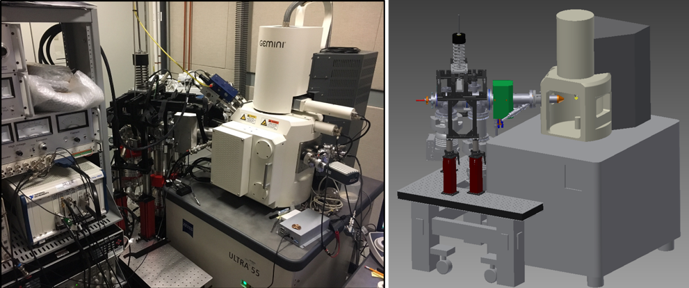 GCIB-SEM prototype constructed at Janelia Research Campus. System consists of an Ionoptika GCIB-10s gun mounted on a Zeiss Ultra SEM.
  GCIB-SEM volume spanning three 1 micron thick sections of fly brain tissue, acquired with 6 x 6 x 4 nm voxels on our GCIB-SEM prototype system.