Main Menu (Mobile)- Block
Main Menu - Block
The Janelia Archives
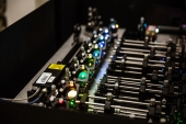
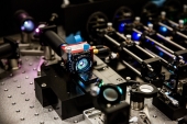
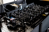
Artifact Name: Lattice Light Sheet Microscope Science
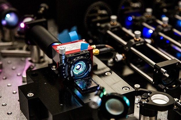
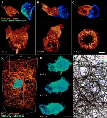
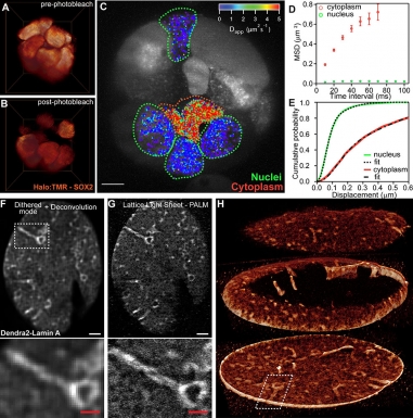
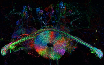
One of the biggest challenges in imaging live cells is observing them without affecting their behavior. Lattice light-sheet microscopy addresses this challenge by using thin sheets of light to illuminate the cell, tissue, or organism one slice at a time, thus reducing the overall exposure to harsh laser light. As a result, the technique is gentle on live samples and has very low phototoxicity/photobleaching effects. Additionally, the use of a plane of illumination light rather than a single point enables faster data acquisition compared to standard confocal or two-photon microscopes. This technology enables researchers to capture previously unseen dynamic biological phenomena in three dimensions, in multiple colors, for multiple hours per experiment.
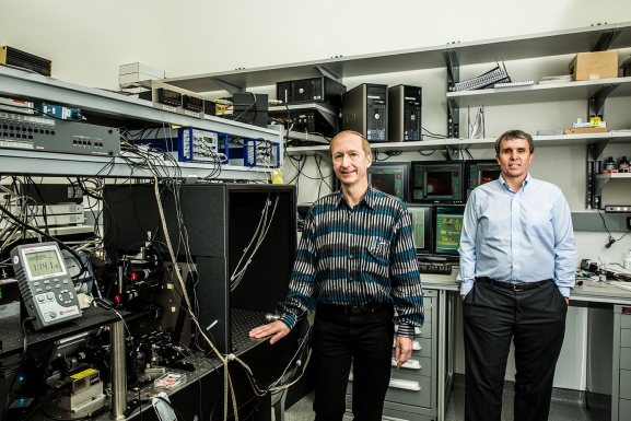
The lattice light-sheet microscope was developed at Janelia by physicist and Nobel Laureate Eric Betzig and his collaborators. Janelia’s philosophy of free dissemination of new technologies has opened up access to the instrument for many researchers around the world. Many outside scientists have benefited from the technique by visiting Janelia through the Advanced Imaging Center, where they used this technology to collect data for their experiments. Some visiting scientists have found the lattice light-sheet microscope so useful that they have subsequently used Betzig’s design plans to build one of their own at their home institutes.