Main Menu (Mobile)- Block
Main Menu - Block
node:field_image_thumbnail | entity_field
custom_misc-custom_misc_featured_summary | block
MouseLight /
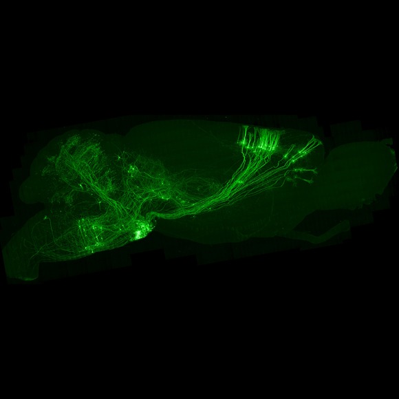
The MouseLight project generates datasets of whole mouse brains imaged at submicron resolution that allows reconstructions of complete axonal arbors of individual neurons across the entire mouse brain.
custom_misc-custom_misc_lab_updates | block
News & Updates
node:field_content_summary | entity_field
An important and challenging goal in modern neuroscience is to understand the brain-wide wiring of neural circuits. To address this, MouseLight is generating axonal collateral maps of individual neurons across the entire mouse brain using high-speed, high-resolution light microscopy.
node:body | entity_field
A major challenge in neuroscience is to relate the structure of neurons to their function. More than 100 years ago, Santiago Ramón y Cajal found that neurons are polarized cells with dendrites and axons, the input and output ends of neurons respectively. His discoveries were based on optical microscopy of histological preparations and single neuron reconstructions. We now know that a neuron's structure is intimately related to its function. Neuronal geometry shapes how synaptic inputs are integrated and predicts a neuron’s position within a circuit. The arborization zones of neuronal axons determine which downstream neurons and brain areas receive information from the neuron, thus forming the basis of circuit organization.
The principal goal of the MouseLight Project is to develop an efficient pipeline to map the complete axonal projections of individual neurons across the entire mouse brain and utilize this platform to generate projection maps of multiple labeled neurons from diverse brain areas. This is a challenging task because on the one hand axons are tiny, with diameters of less than 100 nanometers; tracing axons thus requires resolution and sensitivity at the cutting edge of what is possible with optical microscopy. On the other hand, axonal arbors are huge; individual axons of projection neurons can traverse tens of millimeters across the mouse brain before reaching their targets. We plan to leverage a fast, volumetric serial two-photon microscope designed and developed at Janelia to image the entire mouse brain with submicron spatial resolution, sufficient to resolve and trace individual axons.
To do this we are:
- Evaluating and optimizing sample labeling and clearing techniques necessary for imaging fine structures within three-dimensional volumes of highly scattering tissue.
- Developing computational pipelines for high-resolution axonal reconstructions in the mouse brain.
- Visualizing and annotating whole-brain datasets containing multiple individually labeled neurons
- Collaborating with Janelia Group Leaders to address specific biological questions.
To learn more, please visit our Research Page.
janelia7_blocks-janelia7_block_right_hand_rail | block
"Unfortunately, nature seems unaware of our intellectual need for convenience and unity, and very often takes delight in complication and diversity."
—Santiago Ramón y Cajal, in his 1906 Nobel lecture The structure and connexions of neurons