Main Menu (Mobile)- Block
Main Menu - Block
janelia7_blocks-janelia7_fake_breadcrumb | block
janelia7_blocks-janelia7_support_team_header | block
What We Do
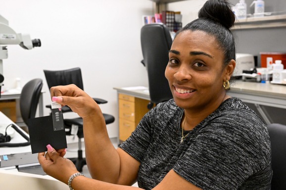
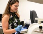
The Anatomy and Histology facility slices tissue and whole organs into sections that are suitable for microscope examination or physiological experiments. You can think of the Histology and Anatomy facility as an extension of scientists’ own labs. Researchers can use the facility’s equipment on their own or utilize the expertise of our on-site staff to request a wide array of services and histological technics. Both routes provide the researchers with the ability to get more science done effectively and efficiently.
node:body | entity_field
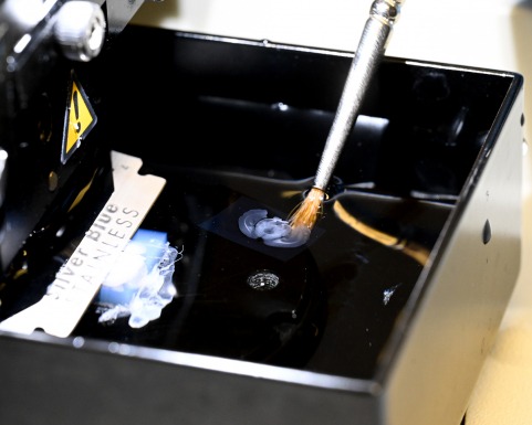
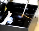
Scientists have access to 1,000 square feet of workspace for tissue fixation, sectioning, immuno-labeling, and tissue expansion of a variety of tissue types. They can tap into staff skilled in antibody-labeling methods, preparing cultured brain slices for physiology experiments, and clearing tissue for optimal imaging. Or they can access a full range of equipment, including two chemical fume hoods for fixatives, vibratomes (4) and cryostats (2), perfusion pumps (5), and microscopes.
Equipment
- Two Leica 3050S cryostats, equipped with CryoJane tape transfer method.
- Four Leica VT1200S vibratomes for sectioning tissue.
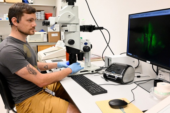
- An Olympus MVX10, a fluorescence dissecting microscope for inspecting endogenous GFP or assessing the quality of antibody labeling in tissue or cells.
- Two non-fluorescent Olympus dissecting scopes for dissecting small tissue.
- Four Watson-Marlow perfusion pumps for tissue fixation.
- SmartBatch+ system for active tissue clearing and antibody labeling.
Services
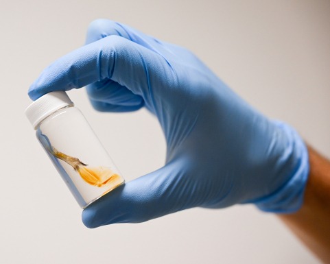
- Anesthesia and perfusion-fixation of mice and rats
- Embedding in OCT and agarose
- 5 µm-thick to 50 µm-thick sections prepared on Leica cryostat.
- 50 um to 1mm thick sections prepared on Leica vibratome.
- Fluorescent antibody labeling and histochemical staining (Nissl, H&E, special stains)
- Preparation of brain slice cultures for physiological recording
- In-situ hybridization- multiple platforms for thin slices (10-20um) and thick slices up to 300um
- Training on vibratomes, cryostats, tissue dissection, and histological techniques
- Imaging- slide scanner and confocal acquisition
- Expansion microscopy sample preparation and imaging
- Whole organ tissue clearing via multiple methods
- Contact us about working with large sample sets
janelia7_blocks-janelia7_block_right_hand_rail | block