Filter
Associated Lab
- Aguilera Castrejon Lab (19) Apply Aguilera Castrejon Lab filter
- Ahrens Lab (75) Apply Ahrens Lab filter
- Aso Lab (42) Apply Aso Lab filter
- Baker Lab (38) Apply Baker Lab filter
- Betzig Lab (116) Apply Betzig Lab filter
- Beyene Lab (15) Apply Beyene Lab filter
- Bock Lab (17) Apply Bock Lab filter
- Branson Lab (56) Apply Branson Lab filter
- Card Lab (43) Apply Card Lab filter
- Cardona Lab (64) Apply Cardona Lab filter
- Chklovskii Lab (13) Apply Chklovskii Lab filter
- Clapham Lab (16) Apply Clapham Lab filter
- Cui Lab (19) Apply Cui Lab filter
- Darshan Lab (12) Apply Darshan Lab filter
- Dennis Lab (3) Apply Dennis Lab filter
- Dickson Lab (46) Apply Dickson Lab filter
- Druckmann Lab (25) Apply Druckmann Lab filter
- Dudman Lab (58) Apply Dudman Lab filter
- Eddy/Rivas Lab (30) Apply Eddy/Rivas Lab filter
- Egnor Lab (11) Apply Egnor Lab filter
- Espinosa Medina Lab (25) Apply Espinosa Medina Lab filter
- Feliciano Lab (16) Apply Feliciano Lab filter
- Fetter Lab (41) Apply Fetter Lab filter
- FIB-SEM Technology (1) Apply FIB-SEM Technology filter
- Fitzgerald Lab (30) Apply Fitzgerald Lab filter
- Freeman Lab (15) Apply Freeman Lab filter
- Funke Lab (46) Apply Funke Lab filter
- Gonen Lab (91) Apply Gonen Lab filter
- Grigorieff Lab (62) Apply Grigorieff Lab filter
- Harris Lab (65) Apply Harris Lab filter
- Heberlein Lab (94) Apply Heberlein Lab filter
- Hermundstad Lab (32) Apply Hermundstad Lab filter
- Hess Lab (80) Apply Hess Lab filter
- Ilanges Lab (4) Apply Ilanges Lab filter
- Jayaraman Lab (49) Apply Jayaraman Lab filter
- Ji Lab (33) Apply Ji Lab filter
- Johnson Lab (7) Apply Johnson Lab filter
- Kainmueller Lab (19) Apply Kainmueller Lab filter
- Karpova Lab (15) Apply Karpova Lab filter
- Keleman Lab (13) Apply Keleman Lab filter
- Keller Lab (77) Apply Keller Lab filter
- Koay Lab (20) Apply Koay Lab filter
- Lavis Lab (162) Apply Lavis Lab filter
- Lee (Albert) Lab (34) Apply Lee (Albert) Lab filter
- Leonardo Lab (23) Apply Leonardo Lab filter
- Li Lab (32) Apply Li Lab filter
- Lippincott-Schwartz Lab (182) Apply Lippincott-Schwartz Lab filter
- Liu (Yin) Lab (9) Apply Liu (Yin) Lab filter
- Liu (Zhe) Lab (65) Apply Liu (Zhe) Lab filter
- Looger Lab (138) Apply Looger Lab filter
- Magee Lab (49) Apply Magee Lab filter
- Menon Lab (18) Apply Menon Lab filter
- Murphy Lab (13) Apply Murphy Lab filter
- O'Shea Lab (8) Apply O'Shea Lab filter
- Otopalik Lab (13) Apply Otopalik Lab filter
- Pachitariu Lab (56) Apply Pachitariu Lab filter
- Pastalkova Lab (19) Apply Pastalkova Lab filter
- Pavlopoulos Lab (19) Apply Pavlopoulos Lab filter
- Pedram Lab (15) Apply Pedram Lab filter
- Podgorski Lab (16) Apply Podgorski Lab filter
- Reiser Lab (55) Apply Reiser Lab filter
- Riddiford Lab (44) Apply Riddiford Lab filter
- Romani Lab (52) Apply Romani Lab filter
- Rubin Lab (149) Apply Rubin Lab filter
- Saalfeld Lab (66) Apply Saalfeld Lab filter
- Satou Lab (18) Apply Satou Lab filter
- Scheffer Lab (38) Apply Scheffer Lab filter
- Schreiter Lab (72) Apply Schreiter Lab filter
- Schulze Lab (1) Apply Schulze Lab filter
- Sgro Lab (23) Apply Sgro Lab filter
- Shroff Lab (35) Apply Shroff Lab filter
- Simpson Lab (23) Apply Simpson Lab filter
- Singer Lab (80) Apply Singer Lab filter
- Spruston Lab (98) Apply Spruston Lab filter
- Stern Lab (160) Apply Stern Lab filter
- Sternson Lab (54) Apply Sternson Lab filter
- Stringer Lab (44) Apply Stringer Lab filter
- Svoboda Lab (136) Apply Svoboda Lab filter
- Tebo Lab (36) Apply Tebo Lab filter
- Tervo Lab (10) Apply Tervo Lab filter
- Tillberg Lab (22) Apply Tillberg Lab filter
- Tjian Lab (64) Apply Tjian Lab filter
- Truman Lab (88) Apply Truman Lab filter
- Turaga Lab (53) Apply Turaga Lab filter
- Turner Lab (38) Apply Turner Lab filter
- Vale Lab (8) Apply Vale Lab filter
- Voigts Lab (5) Apply Voigts Lab filter
- Wang (Meng) Lab (31) Apply Wang (Meng) Lab filter
- Wang (Shaohe) Lab (25) Apply Wang (Shaohe) Lab filter
- Wong-Campos Lab (1) Apply Wong-Campos Lab filter
- Wu Lab (9) Apply Wu Lab filter
- Zlatic Lab (28) Apply Zlatic Lab filter
- Zuker Lab (25) Apply Zuker Lab filter
Associated Project Team
- CellMap (13) Apply CellMap filter
- COSEM (3) Apply COSEM filter
- FIB-SEM Technology (5) Apply FIB-SEM Technology filter
- Fly Descending Interneuron (12) Apply Fly Descending Interneuron filter
- Fly Functional Connectome (14) Apply Fly Functional Connectome filter
- Fly Olympiad (5) Apply Fly Olympiad filter
- FlyEM (56) Apply FlyEM filter
- FlyLight (50) Apply FlyLight filter
- GENIE (47) Apply GENIE filter
- Integrative Imaging (11) Apply Integrative Imaging filter
- Larval Olympiad (2) Apply Larval Olympiad filter
- MouseLight (18) Apply MouseLight filter
- NeuroSeq (1) Apply NeuroSeq filter
- ThalamoSeq (1) Apply ThalamoSeq filter
- Tool Translation Team (T3) (29) Apply Tool Translation Team (T3) filter
- Transcription Imaging (49) Apply Transcription Imaging filter
Publication Date
- 2026 (118) Apply 2026 filter
- 2025 (223) Apply 2025 filter
- 2024 (209) Apply 2024 filter
- 2023 (158) Apply 2023 filter
- 2022 (192) Apply 2022 filter
- 2021 (194) Apply 2021 filter
- 2020 (196) Apply 2020 filter
- 2019 (202) Apply 2019 filter
- 2018 (232) Apply 2018 filter
- 2017 (217) Apply 2017 filter
- 2016 (209) Apply 2016 filter
- 2015 (252) Apply 2015 filter
- 2014 (236) Apply 2014 filter
- 2013 (194) Apply 2013 filter
- 2012 (190) Apply 2012 filter
- 2011 (190) Apply 2011 filter
- 2010 (161) Apply 2010 filter
- 2009 (158) Apply 2009 filter
- 2008 (140) Apply 2008 filter
- 2007 (106) Apply 2007 filter
- 2006 (92) Apply 2006 filter
- 2005 (67) Apply 2005 filter
- 2004 (57) Apply 2004 filter
- 2003 (58) Apply 2003 filter
- 2002 (39) Apply 2002 filter
- 2001 (28) Apply 2001 filter
- 2000 (29) Apply 2000 filter
- 1999 (14) Apply 1999 filter
- 1998 (18) Apply 1998 filter
- 1997 (16) Apply 1997 filter
- 1996 (10) Apply 1996 filter
- 1995 (18) Apply 1995 filter
- 1994 (12) Apply 1994 filter
- 1993 (10) Apply 1993 filter
- 1992 (6) Apply 1992 filter
- 1991 (11) Apply 1991 filter
- 1990 (11) Apply 1990 filter
- 1989 (6) Apply 1989 filter
- 1988 (1) Apply 1988 filter
- 1987 (7) Apply 1987 filter
- 1986 (4) Apply 1986 filter
- 1985 (5) Apply 1985 filter
- 1984 (2) Apply 1984 filter
- 1983 (2) Apply 1983 filter
- 1982 (3) Apply 1982 filter
- 1981 (3) Apply 1981 filter
- 1980 (1) Apply 1980 filter
- 1979 (1) Apply 1979 filter
- 1976 (2) Apply 1976 filter
- 1973 (1) Apply 1973 filter
- 1970 (1) Apply 1970 filter
- 1967 (1) Apply 1967 filter
Type of Publication
4313 Publications
Showing 4151-4160 of 4313 resultsNo abstract available.
The RNA genome of retroviruses is encased within a protein capsid. To gather insight into the assembly and function of this capsid, we used electron cryotomography to image human immunodeficiency virus (HIV) and equine infectious anemia virus (EIAV) particles. While the majority of viral cores appeared closed, a variety of unclosed structures including rolled sheets, extra flaps, and cores with holes in the tip were also seen. Simulations of nonequilibrium growth of elastic sheets recapitulated each of these aberrations and further predicted the occasional presence of seams, for which tentative evidence was also found within the cryotomograms. To test the integrity of viral capsids in vivo, we observed that 25% of cytoplasmic HIV complexes captured by TRIM5α had holes large enough to allow internal green fluorescent protein (GFP) molecules to escape. Together, these findings suggest that HIV assembly at least sometimes involves the union in space of two edges of a curling sheet and results in a substantial number of unclosed forms.
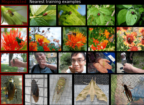
Modern supervised learning algorithms can learn very accurate and complex discriminating functions. But when these classifiers fail, this complexity can also be a drawback because there is no easy, intuitive way to diagnose why they are failing and remedy the problem. This important question has received little attention. To address this problem, we propose a novel method to analyze and understand a classifier's errors. Our method centers around a measure of how much influence a training example has on the classifier's prediction for a test example. To understand why a classifier is mispredicting the label of a given test example, the user can find and review the most influential training examples that caused this misprediction, allowing them to focus their attention on relevant areas of the data space. This will aid the user in determining if and how the training data is inconsistently labeled or lacking in diversity, or if the feature representation is insufficient. As computing the influence of each training example is computationally impractical, we propose a novel distance metric to approximate influence for boosting classifiers that is fast enough to be used interactively. We also show several novel use paradigms of our distance metric. Through experiments, we show that it can be used to find incorrectly or inconsistently labeled training examples, to find specific areas of the data space that need more training data, and to gain insight into which features are missing from the current representation. Code is available at https://github.com/kristinbranson/InfluentialNeighbors.
The eukaryotic core promoter recognition complex was generally thought to play an essential but passive role in the regulation of gene expression. However, recent evidence now indicates that core promoter recognition complexes together with ’non-prototypical’ subunits may have a vital regulatory function in driving cell-specific programmes of transcription during development. Furthermore, new roles for components of these complexes have been identified beyond development; for example, in mediating interactions with chromatin and in maintaining active gene expression across cell divisions.
In practice, understanding the spatial relationships between the surfaces of an object, can significantly improve the performance of object recognition systems. In this paper we propose a novel framework to recognize objects in pictures taken from arbitrary viewpoints. The idea is to maintain the frontal views of the major faces of objects in a global flat map. Then an unfolding warping technique is used to change the pose of the query object in the test view so that all visible surfaces of the object can be observed from a frontal viewpoint, improving the handling of serious occlusions and large viewpoint changes. We demonstrate the effectiveness of our approach through analysis of recognition trials of complex objects with comparison to popular methods.
It is known that sensory deprivation, including postnatal whisker trimming, can lead to severe deficits in the firing rate properties of cortical neurons. Recent results indicate that development of synchronous discharge among cortical neurons is also activity influenced, and that correlated discharge is significantly impaired following loss of bilateral sensory input in rats. Here we investigate whether unilateral whisker trimming (unilateral deprivation or UD) after birth interferes in the same way with the development of synchronous discharge in cortex. We measured the coincidence of spikes among pairs of neurons recorded under urethane anesthesia in one whisker barrel field deprived by trimming all contralateral whiskers for 60 days after birth (UD), and in untrimmed controls (CON). In the septal columns around barrels, UD significantly increased the coincident discharge among cortical neurons compared with CON, most notably in layers II/III. In contrast, synchronous discharge was normal between layer IV UD barrel neurons: i.e., not different from CON. Thus, while bilateral whisker deprivation (BD) produced a global deficit in the development of synchrony in layer IV, UD did not block the development of synchrony between neurons in layer IV barrels and increased synchrony within septal circuits. We conclude that changes in synchronous discharge after UD are unexpectedly different from those recorded after BD, and we speculate that this effect may be due to the driven activity from active commissural inputs arising from the contralateral hemisphere that received normal activity levels during postnatal development.
Compositional generation underlies the systematic and essentially unlimited construction of complex concepts from simpler parts, as is foundational to intelligent behavior, but its underlying neural mechanisms remain unclear. Here we reveal a neural implementation of hierarchical compositional construction of abstract sequences. We demonstrate that in an open-ended setting with very sparse feedback, rats innately utilize hierarchical composition to construct adaptive action sequences that would have been difficult to discover from scratch. Prefrontal neural population representations of these abstract sequences adhere to a low-dimensional format that encodes the orderly progression of elemental units comprising the sequence while converging to a sequence-general endpoint. Higher-level compositions in the hierarchy are systematically related to their lower-level constituent parts, reusing much of the representation, while providing context separation and satisfying format constraints. These neural representations are geometrically identical across animals, pointing to a convergent solution for how knowledge is hierarchically assembled via a compositional mechanism.
Macaque monkeys were tested on a delayed-match-to-multiple-sample task, with either a limited set of well trained images (in randomized sequence) or with never-before-seen images. They performed much better with novel images. False positives were mostly limited to catch-trial image repetitions from the preceding trial. This result implies extremely effective one-shot learning, resembling Standing's finding that people detect familiarity for 10,000 once-seen pictures (with 80% accuracy) (Standing, 1973). Familiarity memory may differ essentially from identification, which embeds and generates contextual information. When encountering another person, we can say immediately whether his or her face is familiar. However, it may be difficult for us to identify the same person. To accompany the psychophysical findings, we present a generic neural network model reproducing these behaviors, based on the same conservative Hebbian synaptic plasticity that generates delay activity identification memory. Familiarity becomes the first step toward establishing identification. Adding an inter-trial reset mechanism limits false positives for previous-trial images. The model, unlike previous proposals, relates repetition-recognition with enhanced neural activity, as recently observed experimentally in 92% of differential cells in prefrontal cortex, an area directly involved in familiarity recognition. There may be an essential functional difference between enhanced responses to novel versus to familiar images: The maximal signal from temporal cortex is for novel stimuli, facilitating additional sensory processing of newly acquired stimuli. The maximal signal for familiar stimuli arising in prefrontal cortex facilitates the formation of selective delay activity, as well as additional consolidation of the memory of the image in an upstream cortical module.
Gaining independent genetic access to discrete cell types is critical to interrogate their biological functions as well as to deliver precise gene therapy. Transcriptomics has allowed us to profile cell populations with extraordinary precision, revealing that cell types are typically defined by a unique combination of genetic markers. Given the lack of adequate tools to target cell types based on multiple markers, most cell types remain inaccessible to genetic manipulation. Here we present CaSSA, a platform to create unlimited genetic switches based on CRISPR/Cas9 (Ca) and the DNA repair mechanism known as single-strand annealing (SSA). CaSSA allows engineering of independent genetic switches, each responding to a specific gRNA. Expressing multiple gRNAs in specific patterns enables multiplex cell-type-specific manipulations and combinatorial genetic targeting. CaSSA is a new genetic tool that conceptually works as an unlimited number of recombinases and will facilitate genetic access to cell types in diverse organisms.