Main Menu (Mobile)- Block
Main Menu - Block

Current Research:
As biologists probe more deeply into tissue, the information content of images degrades due to depth-dependent loss in signal and resolution. The ability to segment structures, count cells, and track motion eventually becomes impossible, preventing further interrogation of biology far from the coverslip.
This image degradation is rooted in distortion of the wavefront of light as it travels through the sample (optical aberrations). Adaptive Optics (AO) promises to reverse these aberrations to enable crisp, bright imaging of structures deep inside living tissues and organisms. However, this tool has not enjoyed broad adoption by the larger biological community because existing approaches to sense wavefront aberrations are slow, expensive, and difficult to use – even by expert microscopists.
Here at Janelia, I work in collaboration with theorists to develop a new computational approach to wavefront sensing that overcomes these barriers and ultimately enable widespread adoption of AO.
Biography
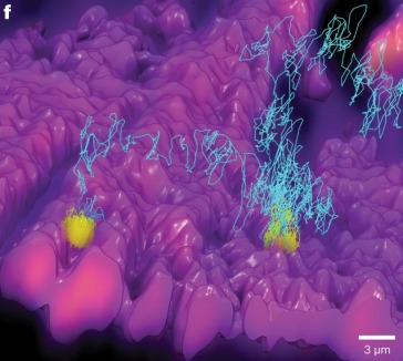
Courtney "CJ" Johnson received her Chemistry Ph.D. from Duke University in Durham, NC, where she learned how to build microscopes from an empty optical table to fully automated operation as a founding member of Kevin Welsher’s lab. During her time at Duke, she spearheaded the design of 3D-FASTR, a new method for 3D Point-Scan Microscopy, and led development of the 3D-TrIm single particle tracking microscope for visualizing high-speed virus-cell interactions in 3D. Before coming to Duke, CJ received her B.S. in Chemistry from Texas Woman’s University in Denton, TX.
Education
Featured Publications