Watching a Synapse Develop in a Young Brain
The Calyx of Held, the largest synapse in the mammalian brain, plays a key role in hearing. The synapse, named because of its resemblance to the whorled calyx of a flower, is the juncture between two important neurons: the neuron that directly receives vibration in the ear, and the postsynaptic neuron that sends the signal to the brain. The connection develops through a process of competition: initially, many different neurons provide inputs to the receiving neuron, but after a couple of days, the competing inputs are weeded out and only one is left. What determines the “winner”?
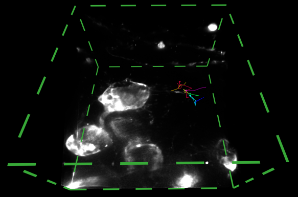
Neuroscientist George Spirou and his research team at West Virginia University study how the Calyx of Held develops and the competition by which a presynaptic cell and postsynaptic cell establish this one-to-one connection. Because brain development happens so fast, a commercially available two-photon microscope, for example, isn’t useful. But the AIC’s cutting-edge lattice light sheet microscope can sample images rapidly enough to watch neurons in a fresh brain slice compete to form a synapse with another neuron. In addition, because the instrument illuminates the sample with a thin light sheet it generates superior image contrast that abrogates the need for powerful laser illumination, thus greatly lowering phototoxicity on the sample. As a result, a biologically active brain slice can be imaged for several hours.
After just two visits, “we were able to observe [pre- and post-synaptic neurons] dance together,” said Spirou. “It is unprecedented to see the early development of synapse formation at such high spatial and temporal resolution.”
About the AIC

The AIC has served as an ideal platform for researchers to test out their novel ideas with emerging microscopy technologies.
Read Our Alumni Stories
Join our Mailing List
Join our mailing list to be notified when calls for proposals open.