node_title | node_title
Equipment
node_body | node_body
We have a full complement of state-of-the-art microscopes and multiple workstations dedicated to image processing. Our imaging capabilities support most experiments involving, but not limited to, fixed and live specimens, cultured cells and tissue, organoids, cleared organs/tissue, zebrafish and drosophila embryos, etc.
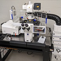
Zeiss LSM 980 with Airyscan (inverted confocal)

- Inverted laser scanning confocal microscope equipped with two multi-Alkali PMTs, 32-channel spectral GaAsP PMTs (up to 13 fps), NIR extended-GaAsP PMT, and AxioCam 506 (monochrome CCD)
- Light sources: 405/445/488/514/561/594/639/730 nm laser lines and Colibri 5 multi-LED illumination system
- Airyscan2 enhanced-resolution capabilities:
- 2x better resolution in all three dimensions
- Improved sensitivity with 4-8x higher SNR
- More gentle excitation (less photobleaching and phototoxicity)
- Multiplexing options with Z-piezo control for imaging up to 8x faster with improved sensitivity
- Spectral unmixing capabilities:
- Acquire 5-8 dyes in parallel with spectral imaging
- Linear unmixing post-acquisition segregates mixed fluorescence signals from dyes and cellular autofluorescence
- Live cell imaging with temperature and CO2 control
- Experiment designer for advanced acquisitions
- Additional features include advanced tiling, photomanipulation, AI Sample Finder, Definite Focus 3, FCS, etc.
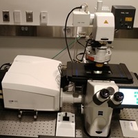
Zeiss LSM 980 NLO with Airyscan (upright confocal)

- Upright confocal and two-photon (2P) laser scanning microscope equipped with two multi-Alkali PMTs, 32-channel spectral GaAsP PMTs (up to 13 fps), NIR extended-GaAsP PMT, BiG-2 non-descanned GaAsP PMT, and AxioCam 506 (monochrome CCD)
- Light sources: 405/445/488/514/561/594/639/730 nm, adjustable 690-1064 nm (Chameleon) laser lines, and X-Cite Xylis illumination system
- Airyscan2 enhanced-resolution capabilities:
- 2x better resolution in all three dimensions
- Improved sensitivity with 4-8x higher SNR
- More gentle excitation (less photobleaching and phototoxicity)
- Multiplexing options with Z-piezo control for imaging up to 8x faster with improved sensitivity
- Spectral unmixing capabilities:
- Acquire 5-8 dyes in parallel with spectral imaging
- Linear unmixing post-acquisition segregates mixed fluorescence signals from dyes and cellular autofluorescence
- Long working distance, water-dipping objectives
- Live cell imaging with temperature and CO2 control
- Experiment designer for advanced acquisitions
- Additional features include advanced tiling, photomanipulation, FCS, etc.
Zeiss LSM 980 (inverted confocal)

- Inverted laser scanning confocal microscope equipped with two multi-Alkali PMTs, 4-channel spectral GaAsP PMTs (up to 13 fps), and AxioCam 506 (monochrome CCD)
- Light sources: 405/445/488/514/561/594/639 nm laser lines and Colibri 5 multi-LED illumination system
- Live cell imaging with temperature and CO2 control
- Experiment designer for advanced acquisitions
- Additional features include advanced tiling, AI Sample Finder, Definite Focus 3, etc.
Zeiss Lightsheet Z.1 (LSFM)

- Sequential dual-side illumination light sheet fluorescence microscope (LSFM) equipped with two liquid cooled pco.edge sCMOS cameras for high sensitivity and speed (up to 100 fps)
- Light sources: 405/445/488/514/561/638 nm laser lines
- 3D acquisition capabilities:
- Thin optical sectioning (light sheet thickness ranges from 2-14 µm) using continuous optical zoom and focusing, dry illumination objectives
- High-speed imaging with very low phototoxicity and photobleaching
- Multiple long working distance, clearing- and water-dipping detection objectives (RI 1.33-1.58)
- Tailored for zebrafish and drosophila embryos, cleared tissue and whole mouse brains, expanded samples, and transparent specimens (up to 20 mm)
- Live cell imaging with temperature and CO2 control
- Additional features include pivot scanning, smart sample holder, tiling, multi-view (360°) imaging, etc.
Zeiss Lightsheet 7 (LSFM)
- Sequential dual-side illumination light sheet fluorescence microscope (LSFM) equipped with two liquid cooled pco.edge sCMOS cameras for high sensitivity and speed (up to 100 fps)
- Light sources: 405/488/561/638 nm laser lines
- 3D acquisition capabilities:
- Thin optical sectioning (light sheet thickness ranges from 2-14 µm) using continuous optical zoom and focusing, dry illumination objectives
- High-speed imaging with very low phototoxicity and photobleaching
- Long working distance, water-dipping detection objective
- Tailored for zebrafish and drosophila embryos, expanded samples, and transparent specimens (up to 20 mm)
- Live cell imaging with temperature and CO2 control
- Additional features include pivot scanning, smart sample holder, tiling, multi-view (360°) imaging, etc.
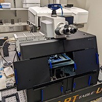
Luxendo MuVi SPIM (LSFM)

- Light sheet fluorescence microscope (LSFM) with horizontal 4-axis arrangement for simultaneous dual-side illumination and dual-side detection equipped with three Hamamatsu ORCA-Flash4.0 V3 sCMOS cameras for high sensitivity and speed (up to 100 fps)
- Light sources: 405/445/488/515/561/642 and 405/488/532 (pulsed) nm laser lines for LSFM and galvo-based photomanipulation/ablation, respectively
- Internal (capillary-based) and external piezo stages for sample translation and rotation
- 3D acquisition capabilities:
- Thin optical sectioning (light sheet thickness ranges from 1.5-8 µm) using motorized beam expander and water-dipping illumination objectives
- High-speed imaging with very low phototoxicity and photobleaching
- Axial waist scanning using TAG lens for uniform illumination across FOV
- Rolling shutter camera detection with synchronization of light sheet scan for improved contrast
- Multiple long working distance, clearing- and water-dipping detection objectives (RI 1.33-1.50)
- Tailored for zebrafish and drosophila embryos, cleared tissue and whole mouse brains, expanded samples, and transparent specimens (up to 20 mm)
- Live cell imaging with temperature and CO2 control
- Additional features include pivot scanning, tiling, multi-view (360°) imaging, photomanipulation/ablation, real-time object tracking, etc.
Lattice Light Sheet (LLSM)
- Two custom-built lattice light sheet microscopes (LLSM) each equipped with two Hamamatsu ORCA-Flash4.0 V3 sCMOS cameras for high sensitivity and speed (up to 100 fps)
- Light sources: 405/488/561/642 nm laser lines
- 3D acquisition capabilities:
- Thin optical sectioning (light sheet thickness ranges from 400-800 nm) with high SNR and optimal resolution of 230 nm (lateral) and 370 nm (axial)
- High-speed imaging with very low phototoxicity and photobleaching
- Long working distance, water-dipping detection objective
- Tailored for cultured cells and tissue, zebrafish and drosophila embryos, expanded samples (ExLLSM), and transparent specimens (up to 4 mm)
- Live cell imaging with temperature control
- Additional features include advanced tiling etc.
Multimodal Optical Scope with Adaptive Imaging Correction (MOSAIC)

- Custom-built instrument combining several imaging modalities for visualizing and quantifying complex biological systems in context with adaptive optics (AO) correction and equipped with two Hamamatsu ORCA-Flash4.0 V3 sCMOS cameras for high sensitivity and speed (up to 100 fps)
- Light sources: 405/445/488/514/560/607/642 and adjustable 680-1080 nm (Chameleon) laser lines
- MOSAIC will offer multiplexed capabilities combined with AO: LLSM with AO (available), ExLLSM (available), 3D SIM, Multifocal image scanning/pixel reassignment, 2P point scanning, 2P Bessel fast functional imaging, 2P Bessel LSFM, 2P Bessel point scanning, Phase imaging, Patterned photostimulation, and 3D single molecule localization
- 3D acquisition capabilities:
- Thin optical sectioning (light sheet thickness ranges from 400-800 nm) with high SNR and optimal resolution of 230 nm (lateral) and 370 nm (axial) as well as extended light sheet imaging tile size (220 µm)
- High-speed imaging with very low phototoxicity and photobleaching
- Multiple long working distance, water-dipping detection objectives
- Simultaneous two-color or sequential multi-color illumination for all imaging modes
- Deformable mirror for AO correction (wavefront sensing) along both excitation and emission light paths for all imaging modes
- Tailored for cultured cells and tissue, zebrafish and drosophila embryos, and transparent specimens
- Volumetric Imaging via Photochemical Sectioning (VIPS) capability:
- 375 nm laser used for cleaving large expanded samples
- VIPS overcomes depth limitations, can be performed in real time, and, unlike physical sectioning, preserves overlapping features for registration.
- Tailored for large tissues expanded in a photodegradable hydrogel
- Live cell imaging with temperature, CO2, and hypoxia/hyperoxia (O2) control
- Additional features include advanced tiling, photomanipulation, etc.
Light sheet-based volumetric Fluorescence Lifetime Imaging Microscopy (vFLIM)

- Custom-built light sheet fluorescence microscope (LSFM) with horizontal 4-axis arrangement for simultaneous dual-side illumination and dual-side detection equipped with a Pi Imaging SPAD 512 camera (up to 16 fps)
- Light sources: 488 and 560 nm pulsed laser lines
- Motorized mirror for dynamic switching between FLIM and Intensity imaging modes
- 3D acquisition capabilities:
- Thin optical sectioning with optimal resolution of 400 nm (lateral) and 2 µm (axial) as well as 200x200 µm FOV
- High-speed imaging with very low phototoxicity and photobleaching
- Sensitivity in the range of 200ps lifetime differences
- Long working distance, water-dipping detection objective
- Tailored for multiplexing, biosensor imaging, molecular environment detection, and tissue-scale force sensing in developing systems
- Live cell imaging with temperature and CO2 control
- Additional features include 'Autopilot' (adaptive imaging framework with 10 degrees of freedom for maintaining optimal spatial resolution and tracking) etc.
CryoSIM

- Custom-built instrument for 3D structured illumination microscopy (SIM) at cryogenic temperatures equipped with a Hamamatsu ORCA-Flash4.0 V3 sCMOS camera for high sensitivity and speed (up to 100 fps)
- Light sources: 488/532/561/642 nm laser lines
- Specifically designed for correlative light-electron microscopy (CLEM) applications with capacity for holding up to five sample coverslips at once
- SIM enhanced-resolution capabilities:
- "True" 3D SIM acquisition (5 phase, 3 angle) generated via a digital SLM resulting in 2x better resolution in all three dimensions
- Uniform intensity 3D SIM images with near-zero photobleaching
- Automated SIM alignment and motorized correction collar optimizes SR image quality for each FOV
- Additional features include advanced tiling (widefield multi-image tiling captures a diffraction-limited image of an entire coverslip automatically in minutes to facilitate sample finding for subsequent SIM and FIB-SEM imaging), temperature/pressure alerts, etc.
Abberior Expert Line STED (inverted confocal / STED / FLIM)

- Inverted confocal microscope equipped with four spectral APDs and monochrome CCD camera
- Light sources: 405/440/485/561/640 and STED 595/775 nm laser lines as well as CoolLED illumination system
- STED super-resolution capabilities:
- Easy3D upgrade for dynamic PSF engineering yields achievable STED resolutions of 25x25 nm (2D) and 80x80x90 nm (3D)
- Tunable lateral and axial resolution (absolute and relative to each other) with aberration correction
- Adaptive illumination for reduced photobleaching
- FLIM capabilities:
- Acquire multiple, spectrally-overlapping and/or weakly fluorescent dyes in parallel
- Immediate segregation of mixed fluorescence signals from dyes and cellular autofluorescence
- Improved STED resolution
- MATRIX detector for improved SNR (background subtraction) and better optical sectioning for 2D-STED
- Deformable mirror for AO correction for all imaging modes
- Live cell imaging with temperature and CO2 control
- Additional features include photomanipulation, Z drift compensation unit, FCS (and STED-FCS), etc.
VisiTech iSIM (inverted array-scanning confocal)

- Inverted array-scanning confocal microscope equipped with a Hamamatsu ORCA-Quest qCMOS camera for high sensitivity and speed (up to 120 fps)
- Light sources: 405/488/561/640 nm laser lines as well as CoolLED illumination system
- iSIM enhanced-resolution capabilities:
- Microlensed array for 2x better spatial resolution
- Real-time resolution improvement that is further improved by deconvolution
- Fast Z-piezo stage
- Multiple high-performance objectives including SR and silicone immersion lenses optimized for live cell imaging
- Live cell imaging with temperature and CO2 control
- Additional features include Z drift compensation unit
Nikon CSU-W1 SoRa with TIRF (inverted spinning disk confocal)

- Inverted spinning disk confocal microscope equipped with one Photometrics Prime 95B 22mm and two Hamamatsu ORCA-Fusion BT sCMOS cameras for high sensitivity and speed (up to 100 fps)
- Light sources: 405/445/488/514/561/594/640 (with Uniformizer) and 488/561/640 and 405/473 nm laser lines for confocal microscopy, TIRF, and galvo-based photomanipulation (PM), respectively, as well as Lumencor Spectra-III LED light engines for DMD-based PM and epifluorescence
- System offers multiplexed capabilities including spinning disk confocal microscopy (50 µm-pinholed disk and SoRa disk), TIRF, and large FOV epifluorescence
- SoRa enhanced-resolution capabilities:
- Microlensed array for 2x better spatial resolution
- Real-time resolution improvement that is further improved by deconvolution
- Fast Z-piezo stage
- Multiple high-performance objectives including SR, TIRF, and silicone immersion lenses optimized for live cell imaging
- Live cell imaging with temperature, CO2, and hypoxia/hyperoxia (O2) control
- JOBS module for advanced acquisitions using High Content Analysis with heat maps representation:
- Create any custom acquisition
- Automated screening of multi-well plates
- Intelligent acquisition capable of modifying settings based on real-time image analysis and feedback
- Additional features include advanced tiling, perfect focus system (PFS), water immersion dispenser, etc.
Nikon CSU-W1 SoRa (inverted spinning disk confocal)
- Inverted spinning disk confocal microscope (50 µm-pinholed disk) equipped with two Hamamatsu ORCA-Fusion BT sCMOS cameras for high sensitivity and speed (up to 100 fps)
- Light sources: 405/488/561/640 nm laser lines as well as Lumencor SOLA light engine
- Fast Z-piezo stage
- Live cell imaging with temperature and CO2 control
- SoRa enhanced-resolution capabilities:
- Microlensed array for 2x better spatial resolution
- Real-time resolution improvement that is further improved by deconvolution
- JOBS module for advanced acquisitions using High Content Analysis with heat maps representation:
- Create any custom acquisition
- Automated screening of multi-well plates
- Intelligent acquisition capable of modifying settings based on real-time image analysis and feedback
- Additional features include advanced tiling, PFS, etc.
Nikon Eclipse Ti (inverted widefield)

- Inverted widefield microscope equipped with an Andor Zyla sCMOS camera for high sensitivity and speed (up to 50 fps)
- Light sources: Lumencor Spectra X LED light engine with multiple filter sets
- Live cell imaging with temperature and CO2 control
- JOBS module for advanced acquisitions using High Content Analysis with heat maps representation:
- Create any custom acquisition
- Automated screening of multi-well plates
- Intelligent acquisition capable of modifying settings based on real-time image analysis and feedback
- Additional features include advanced tiling, PFS, etc.
ASI Ring TIRF

- Custom-built inverted TIRF microscope equipped with a Photometrics Prime 95B camera for high sensitivity and speed (up to 100 fps)
- Light sources: 445/488/561/594/639 nm laser lines
- Improved illumination uniformity using ring TIRF and πShaper
- Fast Z-piezo stage
- Live cell imaging with temperature and CO2 control
- Additional features include CRISP control (autofocus) etc.
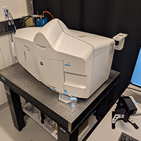
TissueGnostics TissueFAXS 200 (widefield and confocal slide scanner)
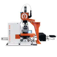
- Combination widefield and spinning disk confocal microscope (50 µm-pinholed disk) operated as a whole slide digital scanner equipped with a Hamamatsu ORCA-Fusion BT sCMOS camera for high sensitivity and speed (up to 100 fps)
- Light sources: Lumencor Celesta (405/446/477/520/546/638/749 nm laser lines) and Spectra X LED light engines with multiple filter sets
- High-throughput capabilities including an autoloader cassette that holds up to 200 slides for unsupervised workflow
- Faster acquisitions are possible by only imaging the tissue sections ("automated" detection)
- Tailored for multi-color slide scanning, tissue cytometry, 4 µm optical sectioning for samples up to 50 µm-thick, and brightfield imaging of chromogenic stained samples
- Additional features include digital stitching of whole slides, tools for cytoplasmic membrane or nuclear measurements, advanced image analysis software, etc.
- Strataquest: Image visualization and analysis software for automated detection/cytometry and context-based quantification tasks across entire slides
Olympus MVX10 (upright macroscope)

- Two upright macro zoom microscopes ("macroscopes") equipped with color CCD cameras
- Light sources: Lumencor SOLA LED light engine with multiple filter sets
- Low and high (NA=0.5) magnification objectives for 50x zoom range without major changes in brightness
- Tailored for dissection, long-term observation of whole organisms, and on-the-fly fluorescence imaging
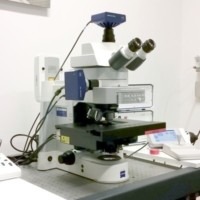
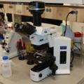
Olympus BX51 (upright compound)
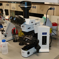
- Upright compound microscope equipped with a 12-bit Olympus DP71 color camera
- 12V/100W halogen illumination source with multiple light filters and objectives including 100x/1.4 oil
- Tailored for color histology/pathology samples and on-the-fly transmitted light viewing
janelia7_blocks-janelia7_block_right_hand_rail | block