The unique system is custom-designed by TissueGnostics (TG) for the Light Microscopy Facility and is currently the only system of its kind.
The TG system combines the efficiency in multi-color slide scanning, confocal sectioning of 5- µm, optical slice for up to 50 µm in sample thickness, and tissue cytometry. In addition, the system is perfectly suited to perform brightfield imaging on chromogenic stained samples.
The system is equipped with TissueFAXS 200 autoloader, which can hold up to 200 slides at once for unsupervised workflow. The system provides seamless stitching of multiple fields of view from large format samples. Images below show the stitched FOVs of a multicolored brain section and the maximal resolution achieved through the system, when the image is fully zoomed in.
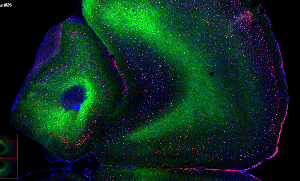
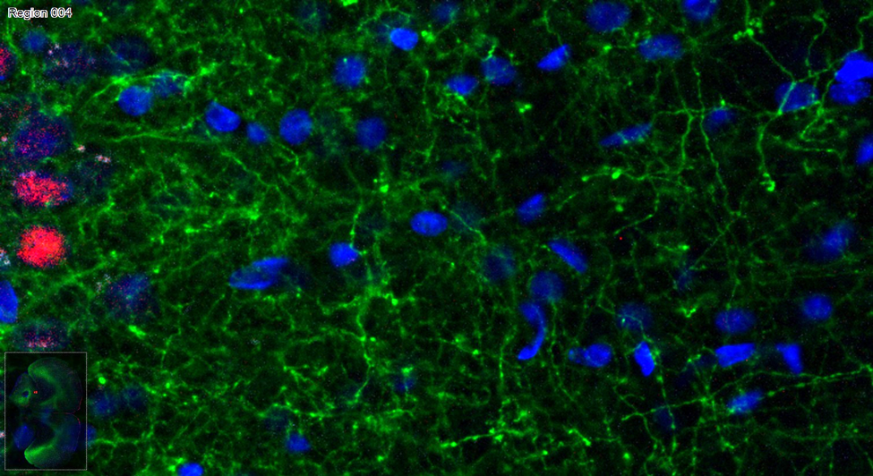
Image analysis using TG’s Strataquest software is context-based. The system can be programmed to perform standard quantification tasks across entire slides. The cytometry capabilities then allow users to customize their quantification strategy by displaying the data in scatterplots that can be further gated or categorized. An example of this can be found here.