How sea urchin larvae build their skeletons
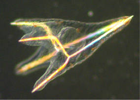
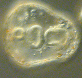
Sea urchin larvae are transparent, delicate, tent-like creatures whose skeletons are made of calcite (a natural form of calcium carbonate crystal) laid down by skeletogenic cells into tube-shaped rods. Smadar Ben Tabou de Leon, of Israel’s University of Haifa, sought to better understand how the skeletal tubes are formed. Previous studies had shown that sea urchin larvae lacking vascular endothelial growth factor (VEGF) – a gene that promotes blood vessel formation in humans – do not form a skeleton, but it wasn’t clear why.
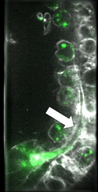
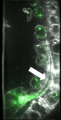
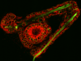
To investigate this question, de Leon needed to observe the skeleton formation in real time, and the AIC’s lattice light sheet microscope had the spatial and temporal resolution suited for the task. Using calcium stain and a membrane marker, she was able to detect and follow the vesicles carrying calcium within the skeletogenic cells as they laid the calcium down in the forming spicule tube. By contrast, the larvae without VEGF receptors also had calcium vesicles in the skeletogenic cells, but they lacked the ability to form the tubular rods and deposit the calcium. “It was a huge jump in our understanding and completely changed how we look at this system,” said de Leon. “It’s a big push forward in our research.”
De Leon was grateful for the support she received at Janelia, in preparing and housing the sea urchins, optimizing the imaging, and later processing the images. “John [Heddleston] and Leong [Chew] were really helpful, both in technical aspects and in designing the experiments,” she said. The brainstorming sessions to discuss how to design rigorous, quantitative imaging experiments were particularly critical to her group’s ultimate success, according to De Leon. “I don’t think we could have made such big progress in such a short time without the assistance and guidance we received from the AIC professionals.”
About the AIC

The AIC has served as an ideal platform for researchers to test out their novel ideas with emerging microscopy technologies.
Read Our Alumni Stories
Join our Mailing List
Join our mailing list to be notified when calls for proposals open.