Watching Intermediate Filaments in Action
Intermediate filaments, a component of the cell cytoskeleton, give the cell its shape and allow it to move. Previously, scientists believed that intermediate filaments were largely stationary. But new work by Vladimir Gelfand, of Northwestern University Feinberg School of Medicine, shows that these filaments are in fact highly mobile and dynamic.
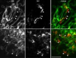
Only 100 nm in diameter, intermediate filaments are tricky to image. So Gelfand has twice visited Janelia’s AIC to use its structured illumination microscope (SIM), which can image macromolecular networks rapidly in detail inside a live cell. Gelfand used special photoconvertible probes to label the intermediate filaments, and leveraged the AIC’s SIM unique capability to perform photoconversion in subcellular regions. Under the microscope, the filaments appear green, but small sections of the filaments can be converted to red, to better track movement of the filaments as a whole.
Using the AIC’s SIM with these probes, Gelfand discovered that, surprisingly, intermediate filaments are extremely dynamic. “In five minutes, they move far away from the area that we initially photoconverted. Nobody had seen that before,” he said. Further imaging studies at the AIC demonstrated that the intermediate filaments move along microtubules. “Intermediate filaments behave like any membrane cargo in the cell. But it’s a non-traditional cargo – it’s not a membrane vesicle or a small protein complex, but a long filament,” he said.
Gelfand pointed out that the staff at the AIC are “as important as the instrument itself.” In particular, he noted that Lin Shao, the application scientist who supported Gelfand during his visit, was “absolutely exceptional. We couldn’t have done this without him.”
About the AIC

The AIC has served as an ideal platform for researchers to test out their novel ideas with emerging microscopy technologies.
Read Our Alumni Stories
Join our Mailing List
Join our mailing list to be notified when calls for proposals open.