Filter
Associated Lab
- Ahrens Lab (1) Apply Ahrens Lab filter
- Aso Lab (1) Apply Aso Lab filter
- Baker Lab (3) Apply Baker Lab filter
- Betzig Lab (7) Apply Betzig Lab filter
- Bock Lab (2) Apply Bock Lab filter
- Branson Lab (1) Apply Branson Lab filter
- Cardona Lab (1) Apply Cardona Lab filter
- Cui Lab (3) Apply Cui Lab filter
- Dickson Lab (2) Apply Dickson Lab filter
- Druckmann Lab (1) Apply Druckmann Lab filter
- Dudman Lab (1) Apply Dudman Lab filter
- Eddy/Rivas Lab (5) Apply Eddy/Rivas Lab filter
- Fetter Lab (4) Apply Fetter Lab filter
- Fitzgerald Lab (1) Apply Fitzgerald Lab filter
- Gonen Lab (5) Apply Gonen Lab filter
- Grigorieff Lab (6) Apply Grigorieff Lab filter
- Heberlein Lab (12) Apply Heberlein Lab filter
- Hermundstad Lab (1) Apply Hermundstad Lab filter
- Hess Lab (2) Apply Hess Lab filter
- Jayaraman Lab (2) Apply Jayaraman Lab filter
- Ji Lab (2) Apply Ji Lab filter
- Kainmueller Lab (1) Apply Kainmueller Lab filter
- Keller Lab (4) Apply Keller Lab filter
- Lavis Lab (5) Apply Lavis Lab filter
- Lee (Albert) Lab (1) Apply Lee (Albert) Lab filter
- Leonardo Lab (2) Apply Leonardo Lab filter
- Lippincott-Schwartz Lab (18) Apply Lippincott-Schwartz Lab filter
- Liu (Zhe) Lab (1) Apply Liu (Zhe) Lab filter
- Looger Lab (7) Apply Looger Lab filter
- Magee Lab (1) Apply Magee Lab filter
- Menon Lab (3) Apply Menon Lab filter
- Murphy Lab (1) Apply Murphy Lab filter
- Pastalkova Lab (1) Apply Pastalkova Lab filter
- Pavlopoulos Lab (2) Apply Pavlopoulos Lab filter
- Reiser Lab (2) Apply Reiser Lab filter
- Riddiford Lab (1) Apply Riddiford Lab filter
- Romani Lab (1) Apply Romani Lab filter
- Rubin Lab (4) Apply Rubin Lab filter
- Satou Lab (3) Apply Satou Lab filter
- Scheffer Lab (2) Apply Scheffer Lab filter
- Schreiter Lab (3) Apply Schreiter Lab filter
- Sgro Lab (2) Apply Sgro Lab filter
- Simpson Lab (3) Apply Simpson Lab filter
- Singer Lab (10) Apply Singer Lab filter
- Spruston Lab (1) Apply Spruston Lab filter
- Stern Lab (4) Apply Stern Lab filter
- Sternson Lab (6) Apply Sternson Lab filter
- Svoboda Lab (7) Apply Svoboda Lab filter
- Tjian Lab (4) Apply Tjian Lab filter
- Truman Lab (1) Apply Truman Lab filter
- Turaga Lab (1) Apply Turaga Lab filter
- Turner Lab (2) Apply Turner Lab filter
- Zlatic Lab (1) Apply Zlatic Lab filter
- Zuker Lab (3) Apply Zuker Lab filter
Associated Project Team
Publication Date
- December 2011 (22) Apply December 2011 filter
- November 2011 (15) Apply November 2011 filter
- October 2011 (14) Apply October 2011 filter
- September 2011 (17) Apply September 2011 filter
- August 2011 (14) Apply August 2011 filter
- July 2011 (10) Apply July 2011 filter
- June 2011 (17) Apply June 2011 filter
- May 2011 (13) Apply May 2011 filter
- April 2011 (11) Apply April 2011 filter
- March 2011 (14) Apply March 2011 filter
- February 2011 (16) Apply February 2011 filter
- January 2011 (27) Apply January 2011 filter
- Remove 2011 filter 2011
Type of Publication
190 Publications
Showing 81-90 of 190 resultsWe designed a real-time computer vision system, the Multi-Worm Tracker (MWT), which can simultaneously quantify the behavior of dozens of Caenorhabditis elegans on a Petri plate at video rates. We examined three traditional behavioral paradigms using this system: spontaneous movement on food, where the behavior changes over tens of minutes; chemotaxis, where turning events must be detected accurately to determine strategy; and habituation of response to tap, where the response is stochastic and changes over time. In each case, manual analysis or automated single-worm tracking would be tedious and time-consuming, but the MWT system allowed rapid quantification of behavior with minimal human effort. Thus, this system will enable large-scale forward and reverse genetic screens for complex behaviors.
Histochemistry (chemistry in the context of biological tissue) is an invaluable set of techniques used to visualize biological structures. This field lies at the interface of organic chemistry, biochemistry, and biology. Integration of these disciplines over the past century has permitted the imaging of cells and tissues using microscopy. Today, by exploiting the unique chemical environments within cells, heterologous expression techniques, and enzymatic activity, histochemical methods can be used to visualize structures in living matter. This review focuses on the labeling techniques and organic fluorophores used in live cells.
HMMER is a software suite for protein sequence similarity searches using probabilistic methods. Previously, HMMER has mainly been available only as a computationally intensive UNIX command-line tool, restricting its use. Recent advances in the software, HMMER3, have resulted in a 100-fold speed gain relative to previous versions. It is now feasible to make efficient profile hidden Markov model (profile HMM) searches via the web. A HMMER web server (http://hmmer.janelia.org) has been designed and implemented such that most protein database searches return within a few seconds. Methods are available for searching either a single protein sequence, multiple protein sequence alignment or profile HMM against a target sequence database, and for searching a protein sequence against Pfam. The web server is designed to cater to a range of different user expertise and accepts batch uploading of multiple queries at once. All search methods are also available as RESTful web services, thereby allowing them to be readily integrated as remotely executed tasks in locally scripted workflows. We have focused on minimizing search times and the ability to rapidly display tabular results, regardless of the number of matches found, developing graphical summaries of the search results to provide quick, intuitive appraisement of them.
Hox genes encode highly conserved transcription factors that regionalize the animal body axis by controlling complex developmental processes. Although they are known to operate in multiple cell types and at different stages, we are still missing the batteries of genes targeted by any one Hox gene over the course of a single developmental process to achieve a particular cell and organ morphology. The transformation of wings into halteres by the Hox gene Ultrabithorax (Ubx) in Drosophila melanogaster presents an excellent model system to study the Hox control of transcriptional networks during successive stages of appendage morphogenesis and cell differentiation. We have used an inducible misexpression system to switch on Ubx in the wing epithelium at successive stages during metamorphosis–in the larva, prepupa, and pupa. We have then used extensive microarray expression profiling and quantitative RT-PCR to identify the primary transcriptional responses to Ubx. We find that Ubx targets range from regulatory genes like transcription factors and signaling components to terminal differentiation genes affecting a broad repertoire of cell behaviors and metabolic reactions. Ubx up- and down-regulates hundreds of downstream genes at each stage, mostly in a subtle manner. Strikingly, our analysis reveals that Ubx target genes are largely distinct at different stages of appendage morphogenesis, suggesting extensive interactions between Hox genes and hormone-controlled regulatory networks to orchestrate complex genetic programs during metamorphosis.
Neural development requires both synapse elaboration and elimination, yet relatively little is known about how these opposing activities are coordinated. Here, we provide evidence Hts/Adducin can serve this function. We show that Drosophila Hts/Adducin is enriched both pre- and postsynaptically at the NMJ. We then demonstrate that presynaptic Hts/Adducin is necessary and sufficient to control two opposing processes associated with synapse remodeling: (1) synapse stabilization as determined by light level and ultrastructural and electrophysiological assays and (2) the elaboration of actin-based, filopodia-like protrusions that drive synaptogenesis and growth. Synapse remodeling is sensitive to Hts/Adducin levels, and we provide evidence that the synaptic localization of Hts/Adducin is controlled via phosphorylation. Mechanistically, Drosophila Hts/Adducin protein has actin-capping activity. We propose that phosphorylation-dependent regulation of Hts/Adducin controls the level, localization, and activity of Hts/Adducin, influencing actin-based synapse elaboration and spectrin-based synapse stabilization. Hts/Adducin may define a mechanism to switch between synapse stability and dynamics.
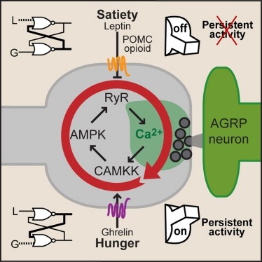
Synaptic plasticity in response to changes in physiologic state is coordinated by hormonal signals across multiple neuronal cell types. Here, we combine cell-type-specific electrophysiological, pharmacological, and optogenetic techniques to dissect neural circuits and molecular pathways controlling synaptic plasticity onto AGRP neurons, a population that regulates feeding. We find that food deprivation elevates excitatory synaptic input, which is mediated by a presynaptic positive feedback loop involving AMP-activated protein kinase. Potentiation of glutamate release was triggered by the orexigenic hormone ghrelin and exhibited hysteresis, persisting for hours after ghrelin removal. Persistent activity was reversed by the anorexigenic hormone leptin, and optogenetic photostimulation demonstrated involvement of opioid release from POMC neurons. Based on these experiments, we propose a memory storage device for physiological state constructed from bistable synapses that are flipped between two sustained activity states by transient exposure to hormones signaling energy levels.
Decoding the wiring diagram of the retina requires simultaneous observation of activity in identified neuron populations. Available recording methods are limited in their scope: electrodes can access only a small fraction of neurons at once, whereas synthetic fluorescent indicator dyes label tissue indiscriminately. Here, we describe a method for studying retinal circuitry at cellular and subcellular levels combining two-photon microscopy and a genetically encoded calcium indicator. Using specific viral and promoter constructs to drive expression of GCaMP3, we labeled all five major neuron classes in the adult mouse retina. Stimulus-evoked GCaMP3 responses as imaged by two-photon microscopy permitted functional cell type annotation. Fluorescence responses were similar to those measured with the small molecule dye OGB-1. Fluorescence intensity correlated linearly with spike rates >10 spikes/s, and a significant change in fluorescence always reflected a significant change in spike firing rate. GCaMP3 expression had no apparent effect on neuronal function. Imaging at subcellular resolution showed compartment-specific calcium dynamics in multiple identified cell types.
Genetically encoded calcium indicators (GECIs), which are based on chimeric fluorescent proteins, can be used to monitor calcium transients in living cells and organisms. Because they are encoded by DNA, GECIs can be delivered to the intact brain noninvasively and targeted to defined populations of neurons and specific subcellular compartments for long-term, repeated measurements in vivo. GECIs have improved iteratively and are becoming useful for imaging neural activity in vivo. Here we summarize extrinsic and intrinsic factors that influence a GECI's performance and provides guidelines for selecting the appropriate GECI for a given application. We also review recent progress in GECI design, optimization, and standardized testing protocols.
We analyze the responses of human observers to an ensemble of monomolecular odorants. Each odorant is characterized by a set of 146 perceptual descriptors obtained from a database of odor character profiles. Each odorant is therefore represented by a point in a highly multidimensional sensory space. In this work we study the arrangement of odorants in this perceptual space. We argue that odorants densely sample a two-dimensional curved surface embedded in the multidimensional sensory space. This surface can account for more than half of the variance of the perceptual data. We also show that only 12% of experimental variance cannot be explained by curved surfaces of substantially small dimensionality (<10). We suggest that these curved manifolds represent the relevant spaces sampled by the human olfactory system, thereby providing surrogates for olfactory sensory space. For the case of 2D approximation, we relate the two parameters on the curved surface to the physico-chemical parameters of odorant molecules. We show that one of the dimensions is related to eigenvalues of molecules’ connectivity matrix, while the other is correlated with measures of molecules’ polarity. We discuss the behavioral significance of these findings.
The basal ganglia play a critical role in the regulation of voluntary action in vertebrates. Our understanding of the function of the basal ganglia relies heavily upon anatomical information, but continued progress will require an understanding of the specific functional roles played by diverse cell types and their connectivity. An increasing number of mouse lines allow extensive identification, characterization, and manipulation of specified cell types in the basal ganglia. Despite the promise of genetically modified mice for elucidating the functional roles of diverse cell types, there is relatively little anatomical data obtained directly in the mouse. Here we have characterized the retrograde labeling obtained from a series of tracer injections throughout the dorsal striatum of adult mice. We found systematic variations in input along both the medial-lateral and anterior-posterior neuraxes in close agreement with canonical features of basal ganglia anatomy in the rat. In addition to the canonical features we have provided experimental support for the importance of non-canonical inputs to the striatum from the raphe nuclei and the amygdala. To look for organization at a finer scale we have analyzed the correlation structure of labeling intensity across our entire dataset. Using this analysis we found substantial local heterogeneity within the large-scale order. From this analysis we conclude that individual striatal sites receive varied combinations of cortical and thalamic input from multiple functional areas, consistent with some earlier studies in the rat that have suggested the presence of a combinatorial map.