Filter
Associated Lab
- Aso Lab (1) Apply Aso Lab filter
- Branson Lab (1) Apply Branson Lab filter
- Card Lab (1) Apply Card Lab filter
- Cardona Lab (1) Apply Cardona Lab filter
- Dickson Lab (1) Apply Dickson Lab filter
- Fetter Lab (2) Apply Fetter Lab filter
- Fitzgerald Lab (1) Apply Fitzgerald Lab filter
- Heberlein Lab (1) Apply Heberlein Lab filter
- Looger Lab (1) Apply Looger Lab filter
- Rubin Lab (3) Apply Rubin Lab filter
- Remove Simpson Lab filter Simpson Lab
- Truman Lab (2) Apply Truman Lab filter
- Zlatic Lab (1) Apply Zlatic Lab filter
Associated Project Team
Publication Date
- 2019 (1) Apply 2019 filter
- 2017 (2) Apply 2017 filter
- 2016 (1) Apply 2016 filter
- 2015 (4) Apply 2015 filter
- 2014 (2) Apply 2014 filter
- 2012 (1) Apply 2012 filter
- 2011 (3) Apply 2011 filter
- 2010 (3) Apply 2010 filter
- 2009 (1) Apply 2009 filter
- 2007 (1) Apply 2007 filter
- 2002 (1) Apply 2002 filter
- 2001 (1) Apply 2001 filter
- 2000 (2) Apply 2000 filter
Type of Publication
23 Publications
Showing 11-20 of 23 results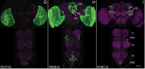
We established a collection of 7,000 transgenic lines of Drosophila melanogaster. Expression of GAL4 in each line is controlled by a different, defined fragment of genomic DNA that serves as a transcriptional enhancer. We used confocal microscopy of dissected nervous systems to determine the expression patterns driven by each fragment in the adult brain and ventral nerve cord. We present image data on 6,650 lines. Using both manual and machine-assisted annotation, we describe the expression patterns in the most useful lines. We illustrate the utility of these data for identifying novel neuronal cell types, revealing brain asymmetry, and describing the nature and extent of neuronal shape stereotypy. The GAL4 lines allow expression of exogenous genes in distinct, small subsets of the adult nervous system. The set of DNA fragments, each driving a documented expression pattern, will facilitate the generation of additional constructs for manipulating neuronal function. synapse was substantially elevated, at the endocytic zone there was no enhanced polymerization activity. We conclude that actin subserves spatially diverse, independently regulated processes throughout spines. Perisynaptic actin forms a uniquely dynamic structure well suited for direct, active regulation of the synapse.
For the overall strategy and methods used to produce the GAL4 lines:
Pfeiffer, B.D., Jenett, A., Hammonds, A.S., Ngo, T.T., Misra, S., Murphy, C., Scully, A., Carlson, J.W., Wan, K.H., Laverty, T.R., Mungall, C., Svirskas, R., Kadonaga, J.T., Doe, C.Q., Eisen, M.B., Celniker, S.E., Rubin, G.M. (2008). Tools for neuroanatomy and neurogenetics in Drosophila. Proc. Natl. Acad. Sci. USA 105, 9715-9720. http://www.pnas.org/content/105/28/9715.full.pdf+html synapse was substantially elevated, at the endocytic zone there was no enhanced polymerization activity. We conclude that actin subserves spatially diverse, independently regulated processes throughout spines. Perisynaptic actin forms a uniquely dynamic structure well suited for direct, active regulation of the synapse.
For data on expression in the embryo:
Manning, L., Purice, M.D., Roberts, J., Pollard, J.L., Bennett, A.L., Kroll, J.R., Dyukareva, A.V., Doan, P.N., Lupton, J.R., Strader, M.E., Tanner, S., Bauer, D., Wilbur, A., Tran, K.D., Laverty, T.R., Pearson, J.C., Crews, S.T., Rubin, G.M., and Doe, C.Q. (2012) Annotated embryonic CNS expression patterns of 5000 GMR GAL4 lines: a resource for manipulating gene expression and analyzing cis-regulatory motifs. Cell Reports (2012) Doi: 10.1016/j.celrep.2012.09.009 http://www.cell.com/cell-reports/fulltext/S2211-1247(12)00290-2 synapse was substantially elevated, at the endocytic zone there was no enhanced polymerization activity. We conclude that actin subserves spatially diverse, independently regulated processes throughout spines. Perisynaptic actin forms a uniquely dynamic structure well suited for direct, active regulation of the synapse.
For data on expression in imaginal discs:
Jory, A., Estella, C., Giorgianni, M.W., Slattery, M., Laverty, T.R., Rubin, G.M., and Mann, R.S. (2012) A survey of 6300 genomic fragments for cis-regulatory activity in the imaginal discs of Drosophila melanogaster. Cell Reports (2012) Doi: 10.1016/j.celrep.2012.09.010 http://www.cell.com/cell-reports/fulltext/S2211-1247(12)00291-4 synapse was substantially elevated, at the endocytic zone there was no enhanced polymerization activity. We conclude that actin subserves spatially diverse, independently regulated processes throughout spines. Perisynaptic actin forms a uniquely dynamic structure well suited for direct, active regulation of the synapse.
For data on expression in the larval nervous system:
Li, H.-H., Kroll, J.R., Lennox, S., Ogundeyi, O., Jeter, J., Depasquale, G., and Truman, J.W. (2013) A GAL4 driver resource for developmental and behavioral studies on the larval CNS of Drosophila. Cell Reports (submitted).
Research in the fruit fly Drosophila melanogaster has led to insights in neural development, axon guidance, ion channel function, synaptic transmission, learning and memory, diurnal rhythmicity, and neural disease that have had broad implications for neuroscience. Drosophila is currently the eukaryotic model organism that permits the most sophisticated in vivo manipulations to address the function of neurons and neuronally expressed genes. Here, we summarize many of the techniques that help assess the role of specific neurons by labeling, removing, or altering their activity. We also survey genetic manipulations to identify and characterize neural genes by mutation, overexpression, and protein labeling. Here, we attempt to acquaint the reader with available options and contexts to apply these methods.
Analyzing Drosophila melanogaster neural expression patterns in thousands of three-dimensional image stacks of individual brains requires registering them into a canonical framework based on a fiducial reference of neuropil morphology. Given a target brain labeled with predefined landmarks, the BrainAligner program automatically finds the corresponding landmarks in a subject brain and maps it to the coordinate system of the target brain via a deformable warp. Using a neuropil marker (the antibody nc82) as a reference of the brain morphology and a target brain that is itself a statistical average of data for 295 brains, we achieved a registration accuracy of 2 μm on average, permitting assessment of stereotypy, potential connectivity and functional mapping of the adult fruit fly brain. We used BrainAligner to generate an image pattern atlas of 2954 registered brains containing 470 different expression patterns that cover all the major compartments of the fly brain.
We developed a multicolor neuron labeling technique in Drosophila melanogaster that combines the power to specifically target different neural populations with the label diversity provided by stochastic color choice. This adaptation of vertebrate Brainbow uses recombination to select one of three epitope-tagged proteins detectable by immunofluorescence. Two copies of this construct yield six bright, separable colors. We used Drosophila Brainbow to study the innervation patterns of multiple antennal lobe projection neuron lineages in the same preparation and to observe the relative trajectories of individual aminergic neurons. Nerve bundles, and even individual neurites hundreds of micrometers long, can be followed with definitive color labeling. We traced motor neurons in the subesophageal ganglion and correlated them to neuromuscular junctions to identify their specific proboscis muscle targets. The ability to independently visualize multiple lineage or neuron projections in the same preparation greatly advances the goal of mapping how neurons connect into circuits.
The role of gamma amino butyric acid (GABA) release and inhibitory neurotransmission in regulating most behaviors remains unclear. The vesicular GABA transporter (VGAT) is required for the storage of GABA in synaptic vesicles and provides a potentially useful probe for inhibitory circuits. However, specific pharmacologic agents for VGAT are not available, and VGAT knockout mice are embryonically lethal, thus precluding behavioral studies. We have identified the Drosophila ortholog of the vesicular GABA transporter gene (which we refer to as dVGAT), immunocytologically mapped dVGAT protein expression in the larva and adult and characterized a dVGAT(minos) mutant allele. dVGAT is embryonically lethal and we do not detect residual dVGAT expression, suggesting that it is either a strong hypomorph or a null. To investigate the function of VGAT and GABA signaling in adult visual flight behavior, we have selectively rescued the dVGAT mutant during development. We show that reduced GABA release does not compromise the active optomotor control of wide-field pattern motion. Conversely, reduced dVGAT expression disrupts normal object tracking and figure-ground discrimination. These results demonstrate that visual behaviors are segregated by the level of GABA signaling in flies, and more generally establish dVGAT as a model to study the contribution of GABA release to other complex behaviors.
The V3D system provides three-dimensional (3D) visualization of gigabyte-sized microscopy image stacks in real time on current laptops and desktops. V3D streamlines the online analysis, measurement and proofreading of complicated image patterns by combining ergonomic functions for selecting a location in an image directly in 3D space and for displaying biological measurements, such as from fluorescent probes, using the overlaid surface objects. V3D runs on all major computer platforms and can be enhanced by software plug-ins to address specific biological problems. To demonstrate this extensibility, we built a V3D-based application, V3D-Neuron, to reconstruct complex 3D neuronal structures from high-resolution brain images. V3D-Neuron can precisely digitize the morphology of a single neuron in a fruitfly brain in minutes, with about a 17-fold improvement in reliability and tenfold savings in time compared with other neuron reconstruction tools. Using V3D-Neuron, we demonstrate the feasibility of building a 3D digital atlas of neurite tracts in the fruitfly brain.
Automatic alignment (registration) of 3D images of adult fruit fly brains is often influenced by the significant displacement of the relative locations of the two optic lobes (OLs) and the center brain (CB). In one of our ongoing efforts to produce a better image alignment pipeline of adult fruit fly brains, we consider separating CB and OLs and align them independently. This paper reports our automatic method to segregate CB and OLs, in particular under conditions where the signal to noise ratio (SNR) is low, the variation of the image intensity is big, and the relative displacement of OLs and CB is substantial. We design an algorithm to find a minimum-cost 3D surface in a 3D image stack to best separate an OL (of one side, either left or right) from CB. This surface is defined as an aggregation of the respective minimum-cost curves detected in each individual 2D image slice. Each curve is defined by a list of control points that best segregate OL and CB. To obtain the locations of these control points, we derive an energy function that includes an image energy term defined by local pixel intensities and two internal energy terms that constrain the curve’s smoothness and length. Gradient descent method is used to optimize this energy function. To improve both the speed and robustness of the method, for each stack, the locations of optimized control points in a slice are taken as the initialization prior for the next slice. We have tested this approach on simulated and real 3D fly brain image stacks and demonstrated that this method can reasonably segregate OLs from CBs despite the aforementioned difficulties.
Drosophila is a marvelous system to study the underlying principles that govern how neural circuits govern behaviors. The scale of the fly brain (approximately 100,000 neurons) and the complexity of the behaviors the fly can perform make it a tractable experimental model organism. In addition, 100 years and hundreds of labs have contributed to an extensive array of tools and techniques that can be used to dissect the function and organization of the fly nervous system. This review discusses both the conceptual challenges and the specific tools for a neurogenetic approach to circuit mapping in Drosophila.
NF-κB signaling has been implicated in neurodegenerative disease, epilepsy, and neuronal plasticity. However, the cellular and molecular activity of NF-κB signaling within the nervous system remains to be clearly defined. Here, we show that the NF-κB and IκB homologs Dorsal and Cactus surround postsynaptic glutamate receptor (GluR) clusters at the Drosophila NMJ. We then show that mutations in dorsal, cactus, and IRAK/pelle kinase specifically impair GluR levels, assayed immunohistochemically and electrophysiologically, without affecting NMJ growth, the size of the postsynaptic density, or homeostatic plasticity. Additional genetic experiments support the conclusion that cactus functions in concert with, rather than in opposition to, dorsal and pelle in this process. Finally, we provide evidence that Dorsal and Cactus act posttranscriptionally, outside the nucleus, to control GluR density. Based upon our data we speculate that Dorsal, Cactus, and Pelle could function together, locally at the postsynaptic density, to specify GluR levels.
The Roundabout (Robo) receptors have been intensively studied for their role in regulating axon guidance in the embryonic nervous system, whereas a role in dendritic guidance has not been explored. In the adult giant fiber system of Drosophila, we have revealed that ectopic Robo expression can regulate the growth and guidance of specific motor neuron dendrites, whereas Robo2 and Robo3 have no effect. We also show that the effect of Robo on dendritic guidance can be suppressed by Commissureless coexpression. Although we confirmed a role for all three Robo receptors in giant fiber axon guidance, the strong axon guidance alterations caused by overexpression of Robo2 or Robo3 have no effect on synaptic connectivity. In contrast, Robo overexpression in the giant fiber seems to directly interfere with synaptic function. We conclude that axon guidance, dendritic guidance, and synaptogenesis are separable processes and that the different Robo family members affect them distinctly.