Filter
Associated Lab
- Ahrens Lab (3) Apply Ahrens Lab filter
- Betzig Lab (8) Apply Betzig Lab filter
- Card Lab (2) Apply Card Lab filter
- Cardona Lab (1) Apply Cardona Lab filter
- Druckmann Lab (1) Apply Druckmann Lab filter
- Eddy/Rivas Lab (2) Apply Eddy/Rivas Lab filter
- Fetter Lab (4) Apply Fetter Lab filter
- Fitzgerald Lab (1) Apply Fitzgerald Lab filter
- Gonen Lab (6) Apply Gonen Lab filter
- Harris Lab (1) Apply Harris Lab filter
- Heberlein Lab (3) Apply Heberlein Lab filter
- Hess Lab (2) Apply Hess Lab filter
- Ji Lab (4) Apply Ji Lab filter
- Kainmueller Lab (1) Apply Kainmueller Lab filter
- Keller Lab (3) Apply Keller Lab filter
- Lavis Lab (4) Apply Lavis Lab filter
- Looger Lab (3) Apply Looger Lab filter
- Magee Lab (4) Apply Magee Lab filter
- Murphy Lab (1) Apply Murphy Lab filter
- Pastalkova Lab (3) Apply Pastalkova Lab filter
- Reiser Lab (1) Apply Reiser Lab filter
- Riddiford Lab (5) Apply Riddiford Lab filter
- Romani Lab (2) Apply Romani Lab filter
- Rubin Lab (4) Apply Rubin Lab filter
- Saalfeld Lab (2) Apply Saalfeld Lab filter
- Satou Lab (1) Apply Satou Lab filter
- Schreiter Lab (3) Apply Schreiter Lab filter
- Sgro Lab (1) Apply Sgro Lab filter
- Shroff Lab (6) Apply Shroff Lab filter
- Spruston Lab (3) Apply Spruston Lab filter
- Stern Lab (4) Apply Stern Lab filter
- Sternson Lab (1) Apply Sternson Lab filter
- Svoboda Lab (6) Apply Svoboda Lab filter
- Tjian Lab (7) Apply Tjian Lab filter
- Truman Lab (2) Apply Truman Lab filter
- Turner Lab (1) Apply Turner Lab filter
- Wu Lab (1) Apply Wu Lab filter
- Zuker Lab (1) Apply Zuker Lab filter
Publication Date
- December 2008 (11) Apply December 2008 filter
- November 2008 (8) Apply November 2008 filter
- October 2008 (7) Apply October 2008 filter
- September 2008 (12) Apply September 2008 filter
- August 2008 (10) Apply August 2008 filter
- July 2008 (14) Apply July 2008 filter
- June 2008 (13) Apply June 2008 filter
- May 2008 (8) Apply May 2008 filter
- April 2008 (7) Apply April 2008 filter
- March 2008 (16) Apply March 2008 filter
- February 2008 (13) Apply February 2008 filter
- January 2008 (21) Apply January 2008 filter
- Remove 2008 filter 2008
Type of Publication
140 Publications
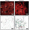
Showing 111-120 of 140 resultsThe identification of synaptic partners is challenging in dense nerve bundles, where many processes occupy regions beneath the resolution of conventional light microscopy. To address this difficulty, we have developed GRASP, a system to label membrane contacts and synapses between two cells in living animals. Two complementary fragments of GFP are expressed on different cells, tethered to extracellular domains of transmembrane carrier proteins. When the complementary GFP fragments are fused to ubiquitous transmembrane proteins, GFP fluorescence appears uniformly along membrane contacts between the two cells. When one or both GFP fragments are fused to synaptic transmembrane proteins, GFP fluorescence is tightly localized to synapses. GRASP marks known synaptic contacts in C. elegans, correctly identifies changes in mutants with altered synaptic specificity, and can uncover new information about synaptic locations as confirmed by electron microscopy. GRASP may prove particularly useful for defining connectivity in complex nervous systems.
We performed patch-clamp recordings from morphologically identified and anatomically mapped pyramidal neurons of the ventral hippocampus to test the hypothesis that bursting neurons are distributed on a gradient from the CA2/CA1 border (proximal) through the subiculum (distal), with more bursting observed at distal locations. We find that the well-defined morphological boundaries between the hippocampal subregions CA1 and subiculum do not correspond to abrupt changes in electrophysiological properties. Rather, we observed that the percentage of bursting neurons is linearly correlated with position in the proximal-distal axis across the CA1 and the subiculum, the percentages of bursting neurons being 10% near the CA1-CA2 border, 24% at the CA1-subiculum border, and higher than 50% in the distal subiculum. The distribution of bursting neurons was paralleled by a gradient in afterdepolarization (ADP) amplitude. We also tested the hypothesis that there was an association between bursting and two previously described morphologically distinct groups of pyramidal neurons (twin and single apical dendrites) in the CA1 region. We found no difference in output mode between single and twin apical dendrite morphologies, which was consistent with the observation that the two morphologies were equally distributed across the transverse axis of the CA1 region. Taken together with the known organization of connections from CA3 to CA1 and CA1 to subiculum, our results indicate that bursting neurons are most likely to be connected to regular spiking neurons and vice versa.
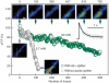
We combined photoactivated localization microscopy (PALM) with live-cell single-particle tracking to create a new method termed sptPALM. We created spatially resolved maps of single-molecule motions by imaging the membrane proteins Gag and VSVG, and obtained several orders of magnitude more trajectories per cell than traditional single-particle tracking enables. By probing distinct subsets of molecules, sptPALM can provide insight into the origins of spatial and temporal heterogeneities in membranes.
Commentary: As a stepping stone to true live cell PALM (see above), our collaborator Jennifer Lippincott-Schwartz suggested using the sparse photoactivation principle of PALM to track the nanoscale motion of thousands of individual molecules within a single living cell. Termed single particle tracking PALM (sptPALM), Jennifer’s postdocs Suliana Manley and Jen Gillette used the method in our PALM rig to create spatially resolved maps of diffusion rates in the plasma membrane of live cells. sptPALM is a powerful tool to study the active cytoskeletal or passive diffusional transport of individual molecules with far more measurements per cell than is possible without sparse photoactivation.
Pulsed lasers are key elements in nonlinear bioimaging techniques such as two-photon fluorescence excitation (TPE) microscopy. Typically, however, only a percent or less of the laser power available can be delivered to the sample before photoinduced damage becomes excessive. Here we describe a passive pulse splitter that converts each laser pulse into a fixed number of sub-pulses of equal energy. We applied the splitter to TPE imaging of fixed mouse brain slices labeled with GFP and show that, in different power regimes, the splitter can be used either to increase the signal rate more than 100-fold or to reduce the rate of photobleaching by over fourfold. In living specimens, the gains were even greater: a ninefold reduction in photobleaching during in vivo imaging of Caenorhabditis elegans larvae, and a six- to 20-fold decrease in the rate of photodamage during calcium imaging of rat hippocampal brain slices.
Pulsed lasers are key elements in nonlinear bioimaging techniques such as two-photon fluorescence excitation (TPE) microscopy. Typically, however, only a percent or less of the laser power available can be delivered to the sample before photoinduced damage becomes excessive. Here we describe a passive pulse splitter that converts each laser pulse into a fixed number of sub-pulses of equal energy. We applied the splitter to TPE imaging of fixed mouse brain slices labeled with GFP and show that, in different power regimes, the splitter can be used either to increase the signal rate more than 100-fold or to reduce the rate of photobleaching by over fourfold. In living specimens, the gains were even greater: a ninefold reduction in photobleaching during in vivo imaging of Caenorhabditis elegans larvae, and a six- to 20-fold decrease in the rate of photodamage during calcium imaging of rat hippocampal brain slices.
Commentary: Na Ji came to me early in her postdoc with an idea to reduce photodamage in nonlinear microscopy by splitting the pulses from an ultrafast laser into multiple subpulses of reduced energy. In six weeks, we constructed a prototype pulse splitter and obtained initial results confirming the validity of her vision. Further experiments with Jeff Magee demonstrated that the splitter could be used to increase imaging speed or reduce photodamage in two photon microscopy by one to two orders of magnitude. This project is a great example of how quickly one can react and exploit new ideas in the Janelia environment.
A miniature, handheld mass spectrometer, based on the rectilinear ion trap mass analyzer, has been applied to air monitoring for traces of toxic compounds. The instrument is battery-operated, hand-portable, and rugged. We anticipate its use in public safety, industrial hygiene, and environmental monitoring. Gaseous samples of nine toxic industrial compounds, phosgene, ethylene oxide, sulfur dioxide, acrylonitrile, cyanogen chloride, hydrogen cyanide, acrolein, formaldehyde, and ethyl parathion, were tested. A sorption trap inlet was constructed to serve as the interface between atmosphere and the vacuum chamber of the mass spectrometer. After selective collection of analytes on the sorbent bed, the sorbent tube was evacuated and then heated to desorb analyte into the instrument. Sampling, detection, identification, and quantitation of all compounds were readily achieved in times of less than 2 min, with detection limits ranging from 800 parts per trillion to 3 parts per million depending on the analyte. For all but one analyte, detection limits were well below (3.5-130 times below) permissible exposure limits. A linear dynamic range of 1-2 orders of magnitude was obtained over the concentration ranges studied (sub-ppbv to ppmv) for all analytes.
Learning and memory has been studied extensively in Drosophila using behavioral, molecular, and genetic approaches. These studies have identified the mushroom body as essential for the formation and retrieval of olfactory memories. We investigated odor responses of the principal neurons of the mushroom body, the Kenyon cells (KCs), in Drosophila using whole cell recordings in vivo. KC responses to odors were highly selective and, thus sparse, compared with those of their direct inputs, the antennal lobe projection neurons (PNs). We examined the mechanisms that might underlie this transformation and identified at least three contributing factors: excitatory synaptic potentials (from PNs) decay rapidly, curtailing temporal integration, PN convergence onto individual KCs is low ( approximately 10 PNs per KC on average), and KC firing thresholds are high. Sparse activity is thought to be useful in structures involved in memory in part because sparseness tends to reduce representation overlaps. By comparing activity patterns evoked by the same odors across olfactory receptor neurons and across KCs, we show that representations of different odors do indeed become less correlated as they progress through the olfactory system.
The fruit fly Drosophila melanogaster performs at least two distinct types of flight initiation. One kind is a stereotyped escape response to a visual stimulus that is mediated by the hard-wired giant fiber neural pathway, and the other is a more variable ;voluntary’ response that can be performed without giant fiber activation. Because the simpler escape take-offs are apparently successful, it is unclear why the fly has multiple pathways to coordinate flight initiation. In this study we use high-speed videography to observe flight initiation in unrestrained wild-type flies and assess the flight performance of each of the two types of take-off. Three-dimensional kinematic analysis of take-off sequences indicates that wing use during the jumping phase of flight initiation is essential for stabilizing flight. During voluntary take-offs, early wing elevation leads to a slower and more stable take-off. In contrast, during visually elicited escapes, the wings are pulled down close to the body during take-off, resulting in tumbling flights in which the fly translates faster but also rotates rapidly about all three of its body axes. Additionally, we find evidence that the power delivered by the legs is substantially greater during visually elicited escapes than during voluntary take-offs. Thus, we find that the two types of Drosophila flight initiation result in different flight performances once the fly is airborne, and that these performances are distinguished by a trade-off between speed and stability.
The evolution of complete metamorphosis in insects is a key innovation that has led to the successful diversification of holometabolous insects, yet the origin of the pupa remains an enigma. Here, we analyzed the expression of the pupal specifier gene broad (br), and the effect on br of isoform-specific, double-stranded RNA-mediated silencing, in a basal holometabolous insect, the beetle Tribolium castaneum. All five isoforms are weakly expressed during the penultimate instar and highly expressed during the prepupal period of the final instar. Application of hydroprene, a juvenile hormone analog, during the penultimate instar caused a repeat of the penultimate br expression patterns, and the formation of supernumerary larvae. Use of dsRNA against the br core region, or against a pair of either the br-Z2 or br-Z3 isoform with the br-Z1 or br-Z4 isoform, produced mobile animals with well-differentiated adult-like appendages, but which retained larval-like urogomphi and epidermis. Disruption of either the br-Z2 or the br-Z3 isoform caused the formation of shorter wings. Disruption of both br-Z1 and br-Z4 caused the appearance of pupal traits in the adults, but disruption of br-Z5 had no morphological effect. Our findings show that the br isoform functions are broadly conserved within the Holometabola and suggest that evolution of br isoform expression may have played an important role in the evolution of the pupa in holometabolous insects.
p-Nitrophenyl acetate is the most commonly used substrate for detecting the catalytic activity of esterases, including those that activate prodrugs in human cells. This substrate is unstable in aqueous solution, limiting its utility. Here, a stable chromogenic substrate for esterases is produced by the structural isolation of an acetyl ester and p-nitroaniline group using a trimethyl lock moiety. Upon ester hydrolysis, unfavorable steric interactions between the three methyl groups of this o-hydroxycinnamic acid derivative encourage rapid lactonization to form a hydrocoumarin and release p-nitroaniline. This "prochromophore" could find use in a variety of assays.