Filter
Associated Lab
- Ahrens Lab (2) Apply Ahrens Lab filter
- Aso Lab (1) Apply Aso Lab filter
- Baker Lab (1) Apply Baker Lab filter
- Branson Lab (1) Apply Branson Lab filter
- Druckmann Lab (3) Apply Druckmann Lab filter
- Harris Lab (3) Apply Harris Lab filter
- Hermundstad Lab (9) Apply Hermundstad Lab filter
- Hess Lab (1) Apply Hess Lab filter
- Remove Jayaraman Lab filter Jayaraman Lab
- Ji Lab (1) Apply Ji Lab filter
- Karpova Lab (1) Apply Karpova Lab filter
- Looger Lab (10) Apply Looger Lab filter
- Podgorski Lab (1) Apply Podgorski Lab filter
- Reiser Lab (2) Apply Reiser Lab filter
- Romani Lab (5) Apply Romani Lab filter
- Rubin Lab (6) Apply Rubin Lab filter
- Saalfeld Lab (1) Apply Saalfeld Lab filter
- Scheffer Lab (1) Apply Scheffer Lab filter
- Schreiter Lab (9) Apply Schreiter Lab filter
- Svoboda Lab (9) Apply Svoboda Lab filter
- Zlatic Lab (1) Apply Zlatic Lab filter
Associated Project Team
Publication Date
- 2025 (1) Apply 2025 filter
- 2024 (3) Apply 2024 filter
- 2023 (1) Apply 2023 filter
- 2022 (3) Apply 2022 filter
- 2021 (1) Apply 2021 filter
- 2020 (4) Apply 2020 filter
- 2019 (4) Apply 2019 filter
- 2018 (3) Apply 2018 filter
- 2017 (4) Apply 2017 filter
- 2016 (3) Apply 2016 filter
- 2015 (4) Apply 2015 filter
- 2014 (1) Apply 2014 filter
- 2013 (3) Apply 2013 filter
- 2012 (2) Apply 2012 filter
- 2011 (2) Apply 2011 filter
- 2010 (2) Apply 2010 filter
- 2009 (2) Apply 2009 filter
- 2007 (1) Apply 2007 filter
- 2006 (1) Apply 2006 filter
- 2003 (1) Apply 2003 filter
Type of Publication
46 Publications
Showing 1-10 of 46 results
Many animals rely on persistent internal representations of continuous variables for working memory, navigation, and motor control. Existing theories typically assume that large networks of neurons are required to maintain such representations accurately; networks with few neurons are thought to generate discrete representations. However, analysis of two-photon calcium imaging data from tethered flies walking in darkness suggests that their small head-direction system can maintain a surprisingly continuous and accurate representation. We thus ask whether it is possible for a small network to generate a continuous, rather than discrete, representation of such a variable. We show analytically that even very small networks can be tuned to maintain continuous internal representations, but this comes at the cost of sensitivity to noise and variations in tuning. This work expands the computational repertoire of small networks, and raises the possibility that larger networks could represent more and higher-dimensional variables than previously thought.
After finding food, a foraging animal must decide whether to continue feeding, or to explore the environment for potentially better options. One strategy to negotiate this tradeoff is to perform local searches around the food but repeatedly return to feed. We studied this behavior in flies and used genetic tools to uncover the underlying mechanisms. Over time, flies gradually expand their search, shifting from primarily exploiting food sources to exploring the environment, a change that is likely driven by increases in satiety. We found that flies’ search patterns preserve these dynamics even as the overall scale of the search is modulated by starvation-induced changes in metabolic state. In contrast, search induced by optogenetic activation of sugar sensing neurons does not show these dynamics. We asked what navigational strategies underlie local search. Using a generative model, we found that a change in locomotor pattern after food consumption could account for repeated returns to the food, but failed to capture relatively direct, long return trajectories. Alternative strategies, such as path integration or sensory taxis could allow flies to return from larger distances. We tested this by individually silencing the fly’s head direction system, olfaction and hygrosensation, and found that the only substantial effect was from perturbing hygrosensation, which reduced the number of long exploratory trips. Our study illustrates that local search is composed of multiple behavioral features that evolve over time based on both internal and external factors, providing a path towards uncovering the underlying neural mechanisms.
Anchoring goals to spatial representations enables flexible navigation but is challenging in novel environments when both representations must be acquired simultaneously. We propose a framework for how Drosophila uses internal representations of head direction (HD) to build goal representations upon selective thermal reinforcement. We show that flies use stochastically generated fixations and directed saccades to express heading preferences in an operant visual learning paradigm and that HD neurons are required to modify these preferences based on reinforcement. We used a symmetric visual setting to expose how flies' HD and goal representations co-evolve and how the reliability of these interacting representations impacts behavior. Finally, we describe how rapid learning of new goal headings may rest on a behavioral policy whose parameters are flexible but whose form is genetically encoded in circuit architecture. Such evolutionarily structured architectures, which enable rapidly adaptive behavior driven by internal representations, may be relevant across species.
Navigating animals continuously integrate velocity signals to update internal representations of their directional heading and spatial location in the environment. How neural circuits combine sensory and motor information to construct these velocity estimates and how these self-motion signals, in turn, update internal representations that support navigational computations are not well understood. Recent work in Drosophila has identified a neural circuit that performs angular path integration to compute the fly's head direction, but the nature of the velocity signal is unknown. Here we identify a pair of neurons necessary for angular path integration that encode the fly's rotational velocity with high accuracy using both visual optic flow and motor information. This estimate of rotational velocity does not rely on a moment-to-moment integration of sensory and motor information. Rather, when visual and motor signals are congruent, these neurons prioritize motor information over visual information, and when the two signals are in conflict, reciprocal inhibition selects either the motor or visual signal. Together, our results suggest that flies update their head direction representation by constructing an estimate of rotational velocity that relies primarily on motor information and only incorporates optic flow signals in specific sensorimotor contexts, such as when the motor signal is absent.
Internal representations are thought to support the generation of flexible, long-timescale behavioral patterns in both animals and artificial agents. Here, we present a novel conceptual framework for how Drosophila use their internal representation of head direction to maintain preferred headings in their surroundings, and how they learn to modify these preferences in the presence of selective thermal reinforcement. To develop the framework, we analyzed flies’ behavior in a classical operant visual learning paradigm and found that they use stochastically generated fixations and directed turns to express their heading preferences. Symmetries in the visual scene used in the paradigm allowed us to expose how flies’ probabilistic behavior in this setting is tethered to their head direction representation. We describe how flies’ ability to quickly adapt their behavior to the rules of their environment may rest on a behavioral policy whose parameters are flexible but whose form is genetically encoded in the structure of their circuits. Many of the mechanisms we outline may also be relevant for rapidly adaptive behavior driven by internal representations in other animals, including mammals.
To flexibly navigate, many animals rely on internal spatial representations that persist when the animal is standing still in darkness, and update accurately by integrating the animal's movements in the absence of localizing sensory cues. Theories of mammalian head direction cells have proposed that these dynamics can be realized in a special class of networks that maintain a localized bump of activity via structured recurrent connectivity, and that shift this bump of activity via angular velocity input. Although there are many different variants of these so-called ring attractor networks, they all rely on large numbers of neurons to generate representations that persist in the absence of input and accurately integrate angular velocity input. Surprisingly, in the fly, Drosophila melanogaster, a head direction representation is maintained by a much smaller number of neurons whose dynamics and connectivity resemble those of a ring attractor network. These findings challenge our understanding of ring attractors and their putative implementation in neural circuits. Here, we analyzed failures of angular velocity integration that emerge in small attractor networks with only a few computational units. Motivated by the peak performance of the fly head direction system in darkness, we mathematically derived conditions under which small networks, even with as few as 4 neurons, achieve the performance of much larger networks. The resulting description reveals that by appropriately tuning the network connectivity, the network can maintain persistent representations over the continuum of head directions, and it can accurately integrate angular velocity inputs. We then analytically determined how performance degrades as the connectivity deviates from this optimally-tuned setting, and we find a trade-off between network size and the tuning precision needed to achieve persistence and accurate integration. This work shows how even small networks can accurately track an animal's movements to guide navigation, and it informs our understanding of the functional capabilities of discrete systems more broadly.
Many animals rely on a representation of head direction for flexible, goal-directed navigation. In insects, a compass-like head direction representation is maintained in a conserved brain region called the central complex. This head direction representation is updated by self-motion information and by tethering to sensory cues in the surroundings through a plasticity mechanism. However, under natural settings, some of these sensory cues may temporarily disappear—for example, when clouds hide the sun—and prominent landmarks at different distances from the insect may move across the animal's field of view during translation, creating potential conflicts for a neural compass. We used two-photon calcium imaging in head-fixed Drosophila behaving in virtual reality to monitor the fly's compass during navigation in immersive naturalistic environments with approachable local landmarks. We found that the fly's compass remains stable even in these settings by tethering to available global cues, likely preserving the animal's ability to perform compass-driven behaviors such as maintaining a constant heading.
Flexible behaviors over long timescales are thought to engage recurrent neural networks in deep brain regions, which are experimentally challenging to study. In insects, recurrent circuit dynamics in a brain region called the central complex (CX) enable directed locomotion, sleep, and context- and experience-dependent spatial navigation. We describe the first complete electron-microscopy-based connectome of the CX, including all its neurons and circuits at synaptic resolution. We identified new CX neuron types, novel sensory and motor pathways, and network motifs that likely enable the CX to extract the fly's head-direction, maintain it with attractor dynamics, and combine it with other sensorimotor information to perform vector-based navigational computations. We also identified numerous pathways that may facilitate the selection of CX-driven behavioral patterns by context and internal state. The CX connectome provides a comprehensive blueprint necessary for a detailed understanding of network dynamics underlying sleep, flexible navigation, and state-dependent action selection.
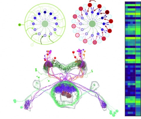
Neural representations of head direction (HD) have been discovered in many species. Theoretical work has proposed that the dynamics associated with these representations are generated, maintained, and updated by recurrent network structures called ring attractors. We evaluated this theorized structure-function relationship by performing electron-microscopy-based circuit reconstruction and RNA profiling of identified cell types in the HD system of Drosophila melanogaster. We identified motifs that have been hypothesized to maintain the HD representation in darkness, update it when the animal turns, and tether it to visual cues. Functional studies provided support for the proposed roles of individual excitatory or inhibitory circuit elements in shaping activity. We also discovered recurrent connections between neuronal arbors with mixed pre- and postsynaptic specializations. Our results confirm that the Drosophila HD network contains the core components of a ring attractor while also revealing unpredicted structural features that might enhance the network's computational power.