Filter
Associated Lab
- Branson Lab (1) Apply Branson Lab filter
- Remove Dudman Lab filter Dudman Lab
- Harris Lab (1) Apply Harris Lab filter
- Hermundstad Lab (1) Apply Hermundstad Lab filter
- Ji Lab (1) Apply Ji Lab filter
- Karpova Lab (3) Apply Karpova Lab filter
- Lavis Lab (2) Apply Lavis Lab filter
- Lee (Albert) Lab (1) Apply Lee (Albert) Lab filter
- Looger Lab (1) Apply Looger Lab filter
- Pachitariu Lab (2) Apply Pachitariu Lab filter
- Spruston Lab (2) Apply Spruston Lab filter
- Sternson Lab (1) Apply Sternson Lab filter
- Svoboda Lab (3) Apply Svoboda Lab filter
- Tervo Lab (2) Apply Tervo Lab filter
- Tillberg Lab (1) Apply Tillberg Lab filter
Associated Project Team
Publication Date
- 2025 (3) Apply 2025 filter
- 2024 (2) Apply 2024 filter
- 2023 (2) Apply 2023 filter
- 2022 (3) Apply 2022 filter
- 2021 (2) Apply 2021 filter
- 2020 (2) Apply 2020 filter
- 2019 (4) Apply 2019 filter
- 2018 (3) Apply 2018 filter
- 2017 (4) Apply 2017 filter
- 2016 (3) Apply 2016 filter
- 2015 (3) Apply 2015 filter
- 2014 (4) Apply 2014 filter
- 2013 (2) Apply 2013 filter
- 2011 (1) Apply 2011 filter
- 2009 (1) Apply 2009 filter
- 2007 (3) Apply 2007 filter
- 2006 (1) Apply 2006 filter
- 2005 (2) Apply 2005 filter
- 2004 (2) Apply 2004 filter
- 2003 (3) Apply 2003 filter
Type of Publication
50 Publications
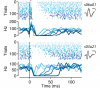
Showing 31-40 of 50 resultsUNLABELLED: Midbrain dopamine (DA) neurons are thought to be a critical node in the circuitry that mediates reward learning. DA neurons receive diverse inputs from regions distributed throughout the neuraxis from frontal neocortex to the mesencephalon. While a great deal is known about changes in the activity of individual DA neurons during learning, much less is known about the functional changes in the microcircuits in which DA neurons are embedded. Here we used local field potentials recorded from the midbrain of behaving mice to show that the midbrain evoked potential (mEP) faithfully reflects the temporal and spatial structure of the phasic response of midbrain neuron populations during conditioning. By comparing the mEP to simultaneously recorded single units, we identified specific components of the mEP that corresponded to phasic DA and non-DA responses to salient stimuli. The DA component of the mEP emerged with the acquisition of a conditioned stimulus, was extinguished following changes in reinforcement contingency, and could be inhibited by pharmacological manipulations that attenuate the phasic responses of DA neurons. In contrast to single-unit recordings, the mEP permitted relatively dense sampling of the midbrain circuit during conditioning and thus could be used to reveal the spatiotemporal structure of multiple intermingled midbrain circuits. Finally, the mEP response was stable for months and thus provides a new approach to study long-term changes in the organization of ventral midbrain microcircuits during learning. SIGNIFICANCE STATEMENT: Neurons that synthesize and release the neurotransmitter dopamine play a critical role in voluntary reward-seeking behavior. Much of our insight into the function of dopamine neurons comes from recordings of individual cells in behaving animals; however, it is notoriously difficult to record from dopamine neurons due to their sparsity and depth, as well as the presence of intermingled non-dopaminergic neurons. Here we show that much of the information that can be learned from recordings of individual dopamine and non-dopamine neurons is also revealed by changes in specific components of the local field potential. This technique provides an accessible measurement that could prove critical to our burgeoning understanding of the molecular, functional, and anatomical diversity of neuron populations in the midbrain.
The basal ganglia plays a significant role in transforming activity in the cerebral cortex into directed behavior, involving motor learning, habit formation and the selection of actions based on desirable outcomes, and the organization of the basal ganglia is intimately linked to that of the cerebral cortex. In this chapter, we focus primarily on the neocortical part of the basal ganglia. A general canonical organizational plan of the neocortical-related basal ganglia is described. An understanding of the canonical organization of the neostriatal part of the basal ganglia, provides a framework for determining the general organizational principles of the parts of the basal ganglia connected with allocortical areas and the amygdala, and this is discussed. While it has been proposed that the basal ganglia provide interactions between disparate functional circuits, another approach might be that there are parallel functional circuits, in which distinct functions are for the most part maintained, or segregated, one from the other. This chapter, however, is biased toward the view that there is maintenance of functional parallel circuits in the organization of the basal ganglia, but that the circuit contains neuroanatomical features that provide for considerable interaction between adjacent circuits.

Compared to the dorsal hippocampus, relatively few studies have characterized neuronal responses in the ventral hippocampus. In particular, it is unclear whether and how cells in the ventral region represent space and/or respond to contextual changes. We recorded from dorsal and ventral CA1 neurons in freely moving mice exposed to manipulations of visuospatial and olfactory contexts. We found that ventral cells respond to alterations of the visuospatial environment such as exposure to novel local cues, cue rotations, and contextual expansion in similar ways to dorsal cells, with the exception of cue rotations. Furthermore, we found that ventral cells responded to odors much more strongly than dorsal cells, particularly to odors of high valence. Similar to earlier studies recording from the ventral hippocampus in CA3, we also found increased scaling of place cell field size along the longitudinal hippocampal axis. Although the increase in place field size observed toward the ventral pole has previously been taken to suggest a decrease in spatial information coded by ventral place cells, we hypothesized that a change in spatial scaling could instead signal a shift in representational coding that preserves the resolution of spatial information. To explore this possibility, we examined population activity using principal component analysis (PCA) and neural location reconstruction techniques. Our results suggest that ventral populations encode a distributed representation of space, and that the resolution of spatial information at the population level is comparable to that of dorsal populations of similar size. Finally, through the use of neural network modeling, we suggest that the redundancy in spatial representation along the longitudinal hippocampal axis may allow the hippocampus to overcome the conflict between memory interference and generalization inherent in neural network memory. Our results suggest that ventral population activity is well suited for generalization across locations and contexts. © 2014 Wiley Periodicals, Inc.
Dysfunction of the basal ganglia produces severe deficits in the timing, initiation, and vigor of movement. These diverse impairments suggest a control system gone awry. In engineered systems, feedback is critical for control. By contrast, models of the basal ganglia highlight feedforward circuitry and ignore intrinsic feedback circuits. In this study, we show that feedback via axon collaterals of substantia nigra projection neurons control the gain of the basal ganglia output. Through a combination of physiology, optogenetics, anatomy, and circuit mapping, we elaborate a general circuit mechanism for gain control in a microcircuit lacking interneurons. Our data suggest that diverse tonic firing rates, weak unitary connections and a spatially diffuse collateral circuit with distinct topography and kinetics from feedforward input is sufficient to implement divisive feedback inhibition. The importance of feedback for engineered systems implies that the intranigral microcircuit, despite its absence from canonical models, could be essential to basal ganglia function. DOI: http://dx.doi.org/10.7554/eLife.02397.001.

A number of recent studies have provided compelling demonstrations that both mice and rats can be trained to perform a variety of behavioral tasks while restrained by mechanical elements mounted to the skull. The independent development of this technique by a number of laboratories has led to diverse solutions. We found that these solutions often used expensive materials and impeded future development and modification in the absence of engineering support. In order to address these issues, here we report on the development of a flexible single hardware design for electrophysiology and imaging both in brain tissue in vitro. Our hardware facilitates the rapid conversion of a single preparation between physiology and imaging system and the conversion of a given system between preparations. In addition, our use of rapid prototyping machines ("3D printers") allows for the deployment of new designs within a day. Here, we present specifications for design and manufacturing as well as some data from our lab demonstrating the suitability of the design for physiology in behaving animals and imaging in vitro and in vivo.

Animals learn both whether and when a reward will occur. Neural models of timing posit that animals learn the mean time until reward perturbed by a fixed relative uncertainty. Nonetheless, animals can learn to perform actions for reward even in highly variable natural environments. Optimal inference in the presence of variable information requires probabilistic models, yet it is unclear whether animals can infer such models for reward timing. Here, we develop a behavioral paradigm in which optimal performance required knowledge of the distribution from which reward delays were chosen. We found that mice were able to accurately adjust their behavior to the SD of the reward delay distribution. Importantly, mice were able to flexibly adjust the amount of prior information used for inference according to the moment-by-moment demands of the task. The ability to infer probabilistic models for timing may allow mice to adapt to complex and dynamic natural environments.

Midbrain dopaminergic (DA) neurons are thought to guide learning via phasic elevations of firing in response to reward predicting stimuli. The mechanism for these signals remains unclear. Using extracellular recording during associative learning, we found that inhibitory neurons in the ventral midbrain of mice responded to salient auditory stimuli with a burst of activity that occurred before the onset of the phasic response of DA neurons. This population of inhibitory neurons exhibited enhanced responses during extinction and was anticorrelated with the phasic response of simultaneously recorded DA neurons. Optogenetic stimulation revealed that this population was, in part, derived from inhibitory projection neurons of the substantia nigra that provide a robust monosynaptic inhibition of DA neurons. Thus, our results elaborate on the dynamic upstream circuits that shape the phasic activity of DA neurons and suggest that the inhibitory microcircuit of the midbrain is critical for new learning in extinction.
The basal ganglia play a critical role in the regulation of voluntary action in vertebrates. Our understanding of the function of the basal ganglia relies heavily upon anatomical information, but continued progress will require an understanding of the specific functional roles played by diverse cell types and their connectivity. An increasing number of mouse lines allow extensive identification, characterization, and manipulation of specified cell types in the basal ganglia. Despite the promise of genetically modified mice for elucidating the functional roles of diverse cell types, there is relatively little anatomical data obtained directly in the mouse. Here we have characterized the retrograde labeling obtained from a series of tracer injections throughout the dorsal striatum of adult mice. We found systematic variations in input along both the medial-lateral and anterior-posterior neuraxes in close agreement with canonical features of basal ganglia anatomy in the rat. In addition to the canonical features we have provided experimental support for the importance of non-canonical inputs to the striatum from the raphe nuclei and the amygdala. To look for organization at a finer scale we have analyzed the correlation structure of labeling intensity across our entire dataset. Using this analysis we found substantial local heterogeneity within the large-scale order. From this analysis we conclude that individual striatal sites receive varied combinations of cortical and thalamic input from multiple functional areas, consistent with some earlier studies in the rat that have suggested the presence of a combinatorial map.
The transformation of synaptic input into patterns of spike output is a fundamental operation that is determined by the particular complement of ion channels that a neuron expresses. Although it is well established that individual ion channel proteins make stochastic transitions between conducting and non-conducting states, most models of synaptic integration are deterministic, and relatively little is known about the functional consequences of interactions between stochastically gating ion channels. Here, we show that a model of stellate neurons from layer II of the medial entorhinal cortex implemented with either stochastic or deterministically gating ion channels can reproduce the resting membrane properties of stellate neurons, but only the stochastic version of the model can fully account for perithreshold membrane potential fluctuations and clustered patterns of spike output that are recorded from stellate neurons during depolarized states. We demonstrate that the stochastic model implements an example of a general mechanism for patterning of neuronal output through activity-dependent changes in the probability of spike firing. Unlike deterministic mechanisms that generate spike patterns through slow changes in the state of model parameters, this general stochastic mechanism does not require retention of information beyond the duration of a single spike and its associated afterhyperpolarization. Instead, clustered patterns of spikes emerge in the stochastic model of stellate neurons as a result of a transient increase in firing probability driven by activation of HCN channels during recovery from the spike afterhyperpolarization. Using this model, we infer conditions in which stochastic ion channel gating may influence firing patterns in vivo and predict consequences of modifications of HCN channel function for in vivo firing patterns.
HCN1 hyperpolarization-activated cation channels act as an inhibitory constraint of both spatial learning and synaptic integration and long-term plasticity in the distal dendrites of hippocampal CA1 pyramidal neurons. However, as HCN1 channels provide an excitatory current, the mechanism of their inhibitory action remains unclear. Here we report that HCN1 channels also constrain CA1 distal dendritic Ca2+ spikes, which have been implicated in the induction of LTP at distal excitatory synapses. Our experimental and computational results indicate that HCN1 channels provide both an active shunt conductance that decreases the temporal integration of distal EPSPs and a tonic depolarizing current that increases resting inactivation of T-type and N-type voltage-gated Ca2+ channels, which contribute to the Ca2+ spikes. This dual mechanism may provide a general means by which HCN channels regulate dendritic excitability.