Filter
Associated Lab
- Aso Lab (1) Apply Aso Lab filter
- Remove Betzig Lab filter Betzig Lab
- Bock Lab (1) Apply Bock Lab filter
- Clapham Lab (2) Apply Clapham Lab filter
- Fetter Lab (2) Apply Fetter Lab filter
- Harris Lab (2) Apply Harris Lab filter
- Hess Lab (6) Apply Hess Lab filter
- Ji Lab (11) Apply Ji Lab filter
- Lavis Lab (8) Apply Lavis Lab filter
- Lippincott-Schwartz Lab (6) Apply Lippincott-Schwartz Lab filter
- Liu (Zhe) Lab (7) Apply Liu (Zhe) Lab filter
- Magee Lab (2) Apply Magee Lab filter
- Rubin Lab (1) Apply Rubin Lab filter
- Saalfeld Lab (2) Apply Saalfeld Lab filter
- Schreiter Lab (1) Apply Schreiter Lab filter
- Shroff Lab (9) Apply Shroff Lab filter
- Singer Lab (1) Apply Singer Lab filter
- Svoboda Lab (2) Apply Svoboda Lab filter
- Tjian Lab (4) Apply Tjian Lab filter
- Turner Lab (1) Apply Turner Lab filter
Associated Project Team
Publication Date
- 2025 (2) Apply 2025 filter
- 2024 (2) Apply 2024 filter
- 2023 (4) Apply 2023 filter
- 2022 (3) Apply 2022 filter
- 2021 (2) Apply 2021 filter
- 2020 (4) Apply 2020 filter
- 2019 (7) Apply 2019 filter
- 2018 (6) Apply 2018 filter
- 2017 (8) Apply 2017 filter
- 2016 (12) Apply 2016 filter
- 2015 (11) Apply 2015 filter
- 2014 (8) Apply 2014 filter
- 2013 (4) Apply 2013 filter
- 2012 (5) Apply 2012 filter
- 2011 (7) Apply 2011 filter
- 2010 (3) Apply 2010 filter
- 2009 (2) Apply 2009 filter
- 2008 (8) Apply 2008 filter
- 2007 (2) Apply 2007 filter
- 2006 (1) Apply 2006 filter
Type of Publication
- Remove Janelia filter Janelia
101 Publications
Showing 41-50 of 101 resultsDespite the apparent simplicity of the xanthene fluorophores, the preparation of caged derivatives with free carboxy groups remains a synthetic challenge. A straightforward and flexible strategy for preparing rhodamine and fluorescein derivatives was developed using reduced, “leuco” intermediates.
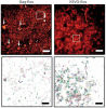
Gap junctions (GJs) represent connexin-rich membrane domains that connect interiors of adjoining cells in mammalian tissues. How fast GJs can respond to bacterial pathogens has not been known previously. Using Bessel beam plane illumination and confocal spinning disk microscopy, we found fast ( 500 ms) formation of connexin-depleted regions (CDRs) inside GJ plaques between cells exposed to AB5 toxins. CDR formation appears as a fast redistribution of connexin channels within GJ plaques with minor changes in outline or geometry. CDR formation does not depend on membrane trafficking or submembrane cytoskeleton and has no effect on GJ conductance. However, CDR responses depend on membrane lipids, can be modified by cholesterol-clustering agents and extracellular K(+) ion concentration, and influence cAMP signaling. The CDR response of GJ plaques to bacterial toxins is a phenomenon observed for all tested connexin isoforms. Through signaling, the CDR response may enable cells to sense exposure to AB5 toxins. CDR formation may reflect lipid-phase separation events in the biological membrane of the GJ plaque, leading to increased connexin packing and lipid reorganization. Our data demonstrate very fast dynamics (in the millisecond-to-second range) within GJ plaques, which previously were considered to be relatively stable, long-lived structures.
Diverse structures facilitate direct exchange of proteins between cells, including plasmadesmata in plants and tunnelling nanotubes in bacteria and higher eukaryotes. Here we describe a new mechanism of protein transfer, flagellar membrane fusion, in the unicellular parasite Trypanosoma brucei. When fluorescently tagged trypanosomes were co-cultured, a small proportion of double-positive cells were observed. The formation of double-positive cells was dependent on the presence of extracellular calcium and was enhanced by placing cells in medium supplemented with fresh bovine serum. Time-lapse microscopy revealed that double-positive cells arose by bidirectional protein exchange in the absence of nuclear transfer. Furthermore, super-resolution microscopy showed that this process occurred in ≤1 minute, the limit of temporal resolution in these experiments. Both cytoplasmic and membrane proteins could be transferred provided they gained access to the flagellum. Intriguingly, a component of the RNAi machinery (Argonaute) was able to move between cells, raising the possibility that small interfering RNAs are transported as cargo. Transmission electron microscopy showed that shared flagella contained two axonemes and two paraflagellar rods bounded by a single membrane. In some cases flagellar fusion was partial and interactions between cells were transient. In other cases fusion occurred along the entire length of the flagellum, was stable for several hours and might be irreversible. Fusion did not appear to be deleterious for cell function: paired cells were motile and could give rise to progeny while fused. The motile flagella of unicellular organisms are related to the sensory cilia of higher eukaryotes, raising the possibility that protein transfer between cells via cilia or flagella occurs more widely in nature.
Actin assembly and inward flow in the plane of the immunological synapse (IS) drives the centralization of T cell receptor microclusters (TCR MCs) and the integrin leukocyte functional antigen 1 (LFA-1). Using structured-illumination microscopy (SIM), we show that actin arcs populating the medial, lamella-like region of the IS arise from linear actin filaments generated by one or more formins present at the IS distal edge. After traversing the outer, Arp2/3-generated, lamellipodia-like region of the IS, these linear filaments are organized by myosin II into antiparallel concentric arcs. Three-dimensional SIM shows that active LFA-1 often aligns with arcs, whereas TCR MCs commonly reside between arcs, and total internal reflection fluorescence SIM shows TCR MCs being swept inward by arcs. Consistently, disrupting actin arc formation via formin inhibition results in less centralized TCR MCs, missegregated integrin clusters, decreased T-B cell adhesion, and diminished TCR signaling. Together, our results define the origin, organization, and functional significance of a major actomyosin contractile structure at the IS that directly propels TCR MC transport.
High-resolution tissue imaging is often compromised by sample-induced optical aberrations that degrade resolution and contrast. While wavefront sensor-based adaptive optics (AO) can measure these aberrations, such hardware solutions are typically complex, expensive to implement, and slow when serially mapping spatially varying aberrations across large fields of view. Here, we introduce AOViFT (Adaptive Optical Vision Fourier Transformer)---a machine learning-based aberration sensing framework built around a 3D multistage Vision Transformer that operates on Fourier domain embeddings. AOViFT infers aberrations and restores diffraction-limited performance in puncta-labeled specimens with substantially reduced computational cost, training time, and memory footprint compared to conventional architectures or real-space networks. We validated AOViFT on live gene-edited zebrafish embryos, demonstrating its ability to correct spatially varying aberrations using either a deformable mirror or post-acquisition deconvolution. By eliminating the need for the guide star and wavefront sensing hardware and simplifying the experimental workflow, AOViFT lowers technical barriers for high-resolution volumetric microscopy across diverse biological samples.
We combined photoactivated localization microscopy (PALM) with live-cell single-particle tracking to create a new method termed sptPALM. We created spatially resolved maps of single-molecule motions by imaging the membrane proteins Gag and VSVG, and obtained several orders of magnitude more trajectories per cell than traditional single-particle tracking enables. By probing distinct subsets of molecules, sptPALM can provide insight into the origins of spatial and temporal heterogeneities in membranes.
Commentary: As a stepping stone to true live cell PALM (see above), our collaborator Jennifer Lippincott-Schwartz suggested using the sparse photoactivation principle of PALM to track the nanoscale motion of thousands of individual molecules within a single living cell. Termed single particle tracking PALM (sptPALM), Jennifer’s postdocs Suliana Manley and Jen Gillette used the method in our PALM rig to create spatially resolved maps of diffusion rates in the plasma membrane of live cells. sptPALM is a powerful tool to study the active cytoskeletal or passive diffusional transport of individual molecules with far more measurements per cell than is possible without sparse photoactivation.
Extending three-dimensional (3D) single-molecule localization microscopy away from the coverslip and into thicker specimens will greatly broaden its biological utility. However, because of the limitations of both conventional imaging modalities and conventional labeling techniques, it is a challenge to localize molecules in three dimensions with high precision in such samples while simultaneously achieving the labeling densities required for high resolution of densely crowded structures. Here we combined lattice light-sheet microscopy with newly developed, freely diffusing, cell-permeable chemical probes with targeted affinity for DNA, intracellular membranes or the plasma membrane. We used this combination to perform high-localization precision, ultrahigh-labeling density, multicolor localization microscopy in samples up to 20 μm thick, including dividing cells and the neuromast organ of a zebrafish embryo. We also demonstrate super-resolution correlative imaging with protein-specific photoactivable fluorophores, providing a mutually compatible, single-platform alternative to correlative light-electron microscopy over large volumes.
In the spindle midzone, microtubules from opposite half-spindles form bundles between segregating chromosomes. Microtubule bundles can either push or restrict chromosome movement during anaphase in different cellular contexts, but how these activities are achieved remains poorly understood. Here, we use high-resolution live-cell imaging to analyze individual microtubule bundles, growing filaments, and chromosome movement in dividing human cells. Within bundles, filament overlap length marked by the cross-linking protein PRC1 decreases during anaphase as chromosome segregation slows. Filament ends within microtubule bundles appear capped despite dynamic PRC1 turnover and submicrometer proximity to growing microtubules. Chromosome segregation distance and rate are increased in two human cell lines when microtubule bundle assembly is prevented via PRC1 knockdown. Upon expressing a mutant PRC1 with reduced microtubule affinity, bundles assemble but chromosome hypersegregation is still observed. We propose that microtubule overlap length reduction, typically linked to pushing forces generated within filament bundles, is needed to properly restrict spindle elongation and position chromosomes within daughter cells.
Neural damage is a devastating outcome of physical trauma. The glia are one of the main effectors of neuronal repair in the nervous system, but the dynamic interactions between peripheral neurons and Schwann cells during injury and regeneration remain incompletely characterized. Here, we combine laser microsurgery, genetic analysis, high-resolution intravital imaging and lattice light-sheet microscopy to study the interaction between Schwann cells and sensory neurons in a zebrafish model of neurotrauma. We found that chronic denervation by neuronal ablation leads to Schwann-cell death, whereas acute denervation by axonal severing does not affect the overall complexity and architecture of the glia. Neuronal-circuit regeneration begins when Schwann cells extend bridging processes to close the injury gap. Regenerating axons grow faster and directionally after the physiological clearing of distal debris by the Schwann cells. This might facilitate circuit repair by ensuring that axons are guided through unoccupied spaces within bands of Büngner towards their original peripheral target. Accordingly, in the absence of Schwann cells, regenerating axons are misrouted, impairing the re-innervation of sensory organs. Our results indicate that regenerating axons use haptotaxis as a directional cue during the reconstitution of a neural circuit. These findings have implications for therapies aimed at neurorepair, which will benefit from preserving the architecture of the peripheral glia during periods of denervation.
Pulsed lasers are key elements in nonlinear bioimaging techniques such as two-photon fluorescence excitation (TPE) microscopy. Typically, however, only a percent or less of the laser power available can be delivered to the sample before photoinduced damage becomes excessive. Here we describe a passive pulse splitter that converts each laser pulse into a fixed number of sub-pulses of equal energy. We applied the splitter to TPE imaging of fixed mouse brain slices labeled with GFP and show that, in different power regimes, the splitter can be used either to increase the signal rate more than 100-fold or to reduce the rate of photobleaching by over fourfold. In living specimens, the gains were even greater: a ninefold reduction in photobleaching during in vivo imaging of Caenorhabditis elegans larvae, and a six- to 20-fold decrease in the rate of photodamage during calcium imaging of rat hippocampal brain slices.