Main Menu (Mobile)- Block
Main Menu - Block
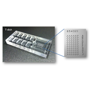
Drosophila Mounting T-Dish
About the Innovation
High-throughput imaging of Drosophila central nervous system specimens requires a precise, standardized arrangement of the samples on a microscope slide. A simple grid makes locating the specimens a straightforward task and streamlines the imaging process. A standardized system of tissue arrangement also guarantees repeatable identification of individual samples.
The Drosophila Mounting T-Dish is a simple device that provides a template for mounting dissected fly nervous systems in a regular array on a glass coverslip. In this device, the coverslip rests on an etched grid of T-shaped marks at the bottom of a saline-filled well. The etched marks help the user place the specimens in standardized positions that quickly allow the imager to locate samples of interest. The marks also help arrange the tissue in the proper orientation to fit an entire adult fly brain into a single 20X microscope field. The mounting dish includes wells to hold specimens in saline until they are mounted and cutouts to ease handling of the coverslip.
The standardized arrangement and orientation of Drosophila nervous systems substantially shortens the time required to acquire high-resolution images. In combination with a slide holder that provides repeatable slide positioning, such as the Janelia Quick-Mount Slide Holder, the T-dish makes it possible to identify individual specimens during multiple imaging sessions correctly. The consistent orientation also minimizes the number of images that researchers must acquire to document an entire nervous system, further reducing the imaging time and the accompanying costs.
The Drosophila Mounting T-dish is designed to mount 80 nervous systems on a 22x22mm coverslip, but modifications for different numbers of specimens and various coverslip sizes are possible.
Advantages:
- Simplifies the regular arrangement of Drosophila CNS samples on a microscope coverslip.
- Improves imaging efficiency.
- Allows reliable identification of individual specimens.
Applications:
- Compatible with many standard imaging techniques, from low-resolution widefield microscopy to high-resolution confocal microscopy.
- Useful in high-throughput, automated microscopy of insect nervous systems or any specimens that benefit from the standardized arrangement on a slide.
Opportunity:
Free to make for Non-Profit Research by downloading designs at Flintbox link to the right.
Rights and designs available for Commercial License.
For inquiries, please reference:
Janelia 2014-037