Main Menu (Mobile)- Block
Main Menu - Block
Kilohertz frame-rate tomographic 2-photon microscope
About the SLAP Microscope
This microscope images megapixel fields of view at over 1000 Hz within scattering tissue by scanning excitation lines at multiple angles and recovering images computationally. It can image large tissue volumes dramatically faster than other 2-photon microscopes.
Figure 1: Rendering of the SLAP Microscope system. A 3D pdf of the system above can be obtained here.
Two-photon microscopes enable high-resolution imaging within scattering tissue, such as the brain. However, conventional point-scanning two-photon microscopes have limited speed, collecting 1-megapixel frames at approximately 16 Hz. The patent-pending Scanned Line Angular Projection Microscope (SLAP Microscope) records > 1-megapixel frames at >1000 Hz.
Rather than recording individual pixels, the SLAP Microscope records tomographic projections by scanning excitation lines within a 2D sample plane at multiple angles. 3D imaging is performed by sequentially scanning 2D planes. High-speed dynamics such as neuronal firing, synaptic activity, sample motion, or particle movement are recovered from tomographic measurements using computational solvers provided with the microscope. As a result, SLAP Microscopy retains the optical sectioning, imaging depth, and resolution benefits of conventional two-photon microscopes.
The SLAP Microscope is a Random Access microscope, allowing user-specified selective excitation of arbitrary patterns within the field of view, as shown in Video 1. Unlike other Random Access microscopes, the frame rate of SLAP is constant regardless of the number of target points or areas imaged. This allows imaging of larger regions, for example, to guard against sample motion without compromising frame rate.
The SLAP microscope uses an inexpensive, powerful femtosecond fiber laser (1030nm fixed wavelength) to provide high-energy pulses for parallel excitation. The Looger, Schreiter, and Podgorski labs have developed fluorescent sensors optimized for this excitation wavelength. For information on sensors or for use at other wavelengths, please contact Kaspar Podgorski.
Video 1: The SLAP Microscope scanning a sample.
System Specifications:
- Imaging speed and Field of View: >1000 Hz at >250 µm diameter FOV
- Number of projection angles: 4
- Imaging depth: >300 µm
- Excitation wavelength: 1030 nm fixed
- Resolution: <0.44 µm FWHM lateral, <1.62 µm FWHM axial @ 1030 nm
Neural and Tissue Imaging Applications:
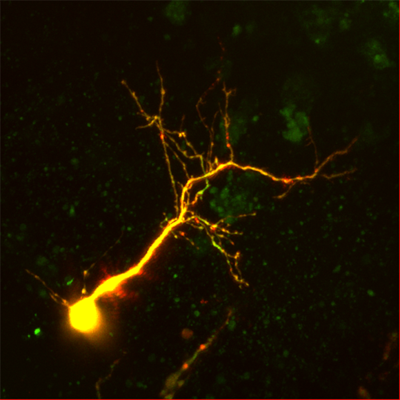
Video 2. In vivo Glutamate Imaging. Recording of dendrites labeled with iGluSnFR.A184V, a glutamate sensor.
Figure 2. Single-neuron optical voltage trace showing evoked action potentials, recorded in vitro with SLAP imaging. The neuron was labeled using Sulfo-RhoVR1 from the Miller Lab, UC Berkeley.
Video 3: Particle Tracking. SLAP 3D tracking of thousands of fluorescent particles flowing through a 250x250x250 micron volume, recorded at over 1.4 billion voxels per second
Figure 3: Submicron precision 3D tracking of brain motion at 1016 Hz using SLAP microscopy.
SLAP imaging includes high-resolution 3D motion-tracking of all samples by comparing tomographic measurements to a reference image stack. Sample position is estimated with high precision (<100nm) and accuracy from individual frames, at >1000 Hz.
Opportunity:
The designs to build an instrument and code for control and analysis are available through a click-through license via the link at the upper right of this page, called "Documentation." The license allows the use of the documentation to produce one SLAP Microscope in your laboratory under the licensee's supervision.
For questions or commercial license inquiries, please contact innovation@janelia.hhmi.org, referencing "SLAP Microscope, tech id: 2017-034."
As with previous microscope designs, Janelia will host training workshops to assemble, align, and use the SLAP Microscope (dates to be determined depending on demand; contact Kaspar Podgorski).
Intellectual Property:
US Patent 10,830,701
See publication for more info.