Main Menu (Mobile)- Block
Main Menu - Block
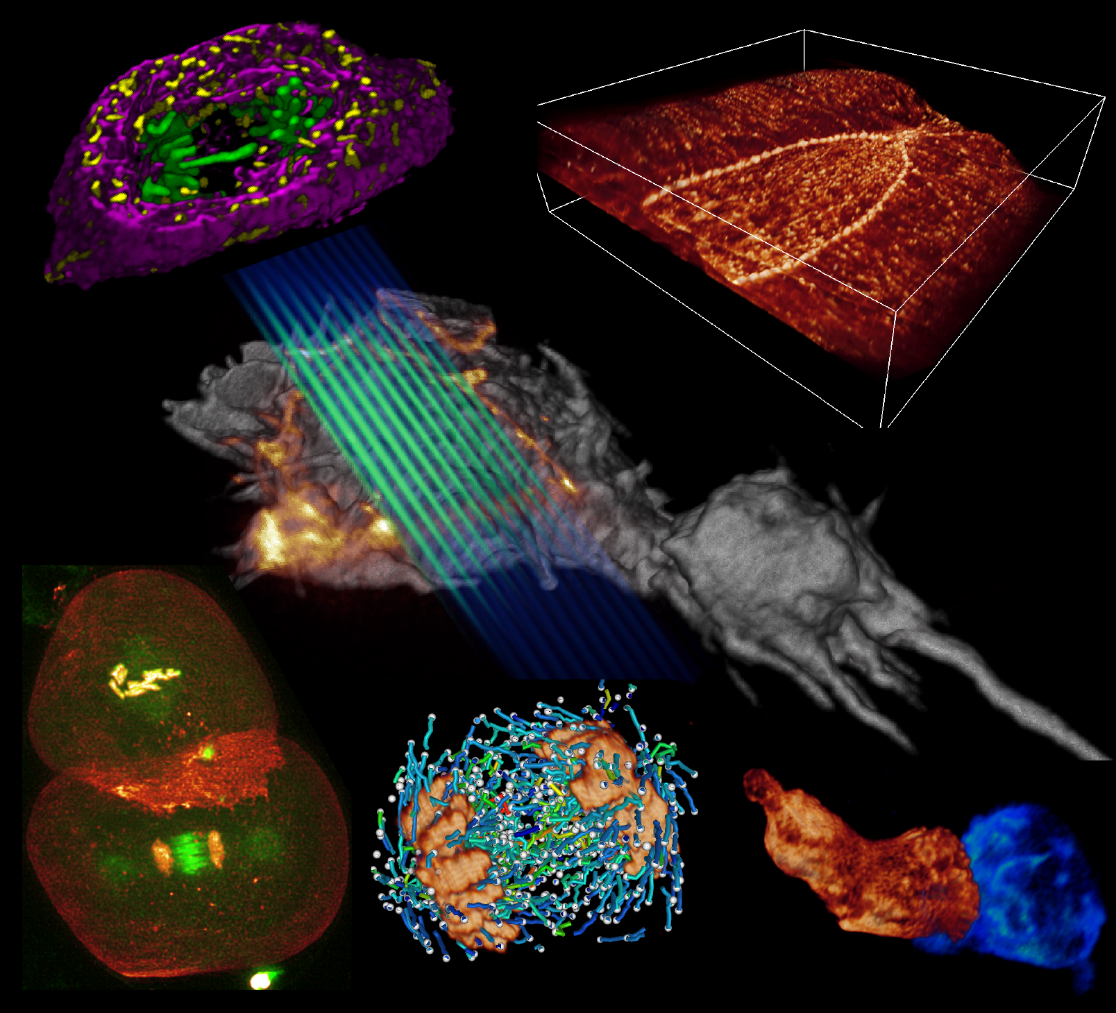
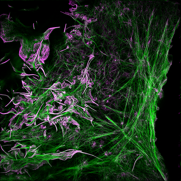
Lattice Light Microscopy
Bessel Beam Plane Illumination Microscopy
The Lattice Light involves using a Bessel beam to illuminate a sample with light sheets that are sufficiently thin to achieve isotropic 3D resolution high-speed 3D fluorescent imaging. As a result, the technology can visualize cellular processes in spatiotemporal detail not attainable with other fluorescent microscopy techniques.
One of the most powerful and widely used imaging modalities in life sciences is fluorescence microscopy. A limitation of fluorescence microscopy is that fluorescent molecules have a fluorescence “budget” (i.e., they can be optically excited for a limited time before they are permanently photobleached). Since both widefield and confocal microscopy excite fluorescence in every plane of the specimen, not just the plane being imaged, they waste the fluorescence budget. They can also be damaging to live specimens. Biological imaging can benefit from a technique that confines excitation to the focal plane to reduce photobleaching and photodamage and reduce out-focus background.
This invention accomplishes that goal by using a Bessel beam to illuminate only the focal plane. The use of the Bessel beam makes this different from previous plane-illumination techniques that have been used in developmental biology. Plane illumination microscopy is limited in practice because the light sheet is not of uniform thickness across the field of view, with the extent of spreading proportional to the width of the field. Where the light sheet is thicker, there is out-of-focus fluorescence excitation, so the benefits of reduced photobleaching are not fully realized. A Bessel beam is a non-diffracting and self-correcting beam that is not subject to this widening and is uniformly thin.
This technology has been demonstrated in conjunction with structured illumination and two-photon microscopy to provide 3D isotropic resolution of ~0.3 µm and speeds up to nearly 200 image planes per second. It can generate very thin (<0.5µm) sheets well suited for 3D subcellular imaging. It has captured dynamic images of mitochondria, intracellular vesicles, and mitotic chromosomes in live cells.
Advantages:
- Achieves visualization of cellular processes in four-dimensional spatiotemporal detail that cannot be attained by other existing fluorescence microscopy techniques
- Offers 3D isotropic resolution to 0.3 µm and imaging speeds up to nearly 200 image planes per second.
- Reduced out-of-focus haze, superior axial resolution, and substantially reduced photobleaching, compared to DSLM and confocal microscopy.
Applications:
- High-resolution imaging of both fixed and living cells
- Long term imaging of dynamic intracellular processes
Publication:
Planchon, T., Betzig, E., et al., Rapid three-dimensional isotropic imaging of living cells using Bessel beam plane illumination. Nature Methods, doi:10.1038/nmeth.1586 (2011)
Patents Status:
Issued U.S. Patents 8,711,211, 9,448,395, 9,223,125, 9,477,074, 9,791,685, 9,891,421, 10,509,217, 10,051,240 and 10,721,441
Opportunity:
Free for Non-Profit Research - you can request a Research License by emailing innovation@janelia.hhmi.org. A research license allows you to practice the invention and access the confidential Lattice Light microscope documentation.
For inquiries, please reference:
Janelia 2009-005 and 2012-010