Filter
Associated Lab
- Ahrens Lab (2) Apply Ahrens Lab filter
- Betzig Lab (1) Apply Betzig Lab filter
- Beyene Lab (1) Apply Beyene Lab filter
- Branson Lab (1) Apply Branson Lab filter
- Darshan Lab (1) Apply Darshan Lab filter
- Dickson Lab (3) Apply Dickson Lab filter
- Druckmann Lab (2) Apply Druckmann Lab filter
- Dudman Lab (3) Apply Dudman Lab filter
- Fitzgerald Lab (3) Apply Fitzgerald Lab filter
- Gonen Lab (1) Apply Gonen Lab filter
- Harris Lab (2) Apply Harris Lab filter
- Heberlein Lab (1) Apply Heberlein Lab filter
- Hermundstad Lab (2) Apply Hermundstad Lab filter
- Hess Lab (3) Apply Hess Lab filter
- Jayaraman Lab (6) Apply Jayaraman Lab filter
- Lavis Lab (1) Apply Lavis Lab filter
- Lee (Albert) Lab (2) Apply Lee (Albert) Lab filter
- Lippincott-Schwartz Lab (1) Apply Lippincott-Schwartz Lab filter
- Looger Lab (2) Apply Looger Lab filter
- Pachitariu Lab (1) Apply Pachitariu Lab filter
- Podgorski Lab (1) Apply Podgorski Lab filter
- Reiser Lab (1) Apply Reiser Lab filter
- Romani Lab (1) Apply Romani Lab filter
- Rubin Lab (1) Apply Rubin Lab filter
- Saalfeld Lab (1) Apply Saalfeld Lab filter
- Scheffer Lab (1) Apply Scheffer Lab filter
- Schreiter Lab (4) Apply Schreiter Lab filter
- Spruston Lab (2) Apply Spruston Lab filter
- Stringer Lab (2) Apply Stringer Lab filter
- Svoboda Lab (4) Apply Svoboda Lab filter
- Turner Lab (1) Apply Turner Lab filter
Associated Project Team
Associated Support Team
- Anatomy and Histology (2) Apply Anatomy and Histology filter
- Invertebrate Shared Resource (6) Apply Invertebrate Shared Resource filter
- Remove Janelia Experimental Technology filter Janelia Experimental Technology
- Molecular Genomics (3) Apply Molecular Genomics filter
- Project Technical Resources (4) Apply Project Technical Resources filter
- Scientific Computing Software (3) Apply Scientific Computing Software filter
- Scientific Computing Systems (2) Apply Scientific Computing Systems filter
- Viral Tools (2) Apply Viral Tools filter
Publication Date
- 2025 (3) Apply 2025 filter
- 2024 (1) Apply 2024 filter
- 2023 (3) Apply 2023 filter
- 2022 (3) Apply 2022 filter
- 2021 (2) Apply 2021 filter
- 2020 (8) Apply 2020 filter
- 2019 (6) Apply 2019 filter
- 2018 (4) Apply 2018 filter
- 2016 (2) Apply 2016 filter
- 2015 (2) Apply 2015 filter
- 2014 (2) Apply 2014 filter
- 2010 (1) Apply 2010 filter
37 Janelia Publications
Showing 11-20 of 37 resultsTumors are complex ecosystems composed of malignant and non-malignant cells embedded in a dynamic extracellular matrix (ECM). In the tumor microenvironment, molecular phenotypes are controlled by cell-cell and ECM interactions in 3D cellular neighborhoods (CNs). While their inhibition can impede tumor progression, routine molecular tumor profiling fails to capture cellular interactions. Single-cell spatial transcriptomics (ST) maps receptor-ligand interactions but usually remains limited to 2D tissue sections and lacks ECM readouts. Here, we integrate 3D ST with ECM imaging in serial sections from one clinical lung carcinoma to systematically quantify molecular states, cell-cell interactions, and ECM remodeling in CN. Our integrative analysis pinpointed known immune escape and tumor invasion mechanisms, revealing several druggable drivers of tumor progression in the patient under study. This proof-of-principle study highlights the potential of in-depth CN profiling in routine clinical samples to inform microenvironment-directed therapies. A record of this paper's transparent peer review process is included in the supplemental information.
The interplay between two major forebrain structures - cortex and subcortical striatum - is critical for flexible, goal-directed action. Traditionally, it has been proposed that striatum is critical for selecting what type of action is initiated while the primary motor cortex is involved in the online control of movement execution. Recent data indicates that striatum may also be critical for specifying movement execution. These alternatives have been difficult to reconcile because when comparing very distinct actions, as in the vast majority of work to date, they make essentially indistinguishable predictions. Here, we develop quantitative models to reveal a somewhat paradoxical insight: only comparing neural activity during similar actions makes strongly distinguishing predictions. We thus developed a novel reach-to-pull task in which mice reliably selected between two similar, but distinct reach targets and pull forces. Simultaneous cortical and subcortical recordings were uniquely consistent with a model in which cortex and striatum jointly specify flexible parameters of action during movement execution.
Within cells, the spatial compartmentalization of thousands of distinct proteins serves a multitude of diverse biochemical needs. Correlative super-resolution (SR) fluorescence and electron microscopy (EM) can elucidate protein spatial relationships to global ultrastructure, but has suffered from tradeoffs of structure preservation, fluorescence retention, resolution, and field of view. We developed a platform for three-dimensional cryogenic SR and focused ion beam-milled block-face EM across entire vitreously frozen cells. The approach preserves ultrastructure while enabling independent SR and EM workflow optimization. We discovered unexpected protein-ultrastructure relationships in mammalian cells including intranuclear vesicles containing endoplasmic reticulum-associated proteins, web-like adhesions between cultured neurons, and chromatin domains subclassified on the basis of transcriptional activity. Our findings illustrate the value of a comprehensive multimodal view of ultrastructural variability across whole cells.
The motor cortex controls skilled arm movement by sending temporal patterns of activity to lower motor centres. Local cortical dynamics are thought to shape these patterns throughout movement execution. External inputs have been implicated in setting the initial state of the motor cortex, but they may also have a pattern-generating role. Here we dissect the contribution of local dynamics and inputs to cortical pattern generation during a prehension task in mice. Perturbing cortex to an aberrant state prevented movement initiation, but after the perturbation was released, cortex either bypassed the normal initial state and immediately generated the pattern that controls reaching or failed to generate this pattern. The difference in these two outcomes was probably a result of external inputs. We directly investigated the role of inputs by inactivating the thalamus; this perturbed cortical activity and disrupted limb kinematics at any stage of the movement. Activation of thalamocortical axon terminals at different frequencies disrupted cortical activity and arm movement in a graded manner. Simultaneous recordings revealed that both thalamic activity and the current state of cortex predicted changes in cortical activity. Thus, the pattern generator for dexterous arm movement is distributed across multiple, strongly interacting brain regions.
Behavioral strategies employed for chemotaxis have been described across phyla, but the sensorimotor basis of this phenomenon has seldom been studied in naturalistic contexts. Here, we examine how signals experienced during free olfactory behaviors are processed by first-order olfactory sensory neurons (OSNs) of the Drosophila larva. We find that OSNs can act as differentiators that transiently normalize stimulus intensity-a property potentially derived from a combination of integral feedback and feed-forward regulation of olfactory transduction. In olfactory virtual reality experiments, we report that high activity levels of the OSN suppress turning, whereas low activity levels facilitate turning. Using a generalized linear model, we explain how peripheral encoding of olfactory stimuli modulates the probability of switching from a run to a turn. Our work clarifies the link between computations carried out at the sensory periphery and action selection underlying navigation in odor gradients.
Internal representations are thought to support the generation of flexible, long-timescale behavioral patterns in both animals and artificial agents. Here, we present a novel conceptual framework for how Drosophila use their internal representation of head direction to maintain preferred headings in their surroundings, and how they learn to modify these preferences in the presence of selective thermal reinforcement. To develop the framework, we analyzed flies’ behavior in a classical operant visual learning paradigm and found that they use stochastically generated fixations and directed turns to express their heading preferences. Symmetries in the visual scene used in the paradigm allowed us to expose how flies’ probabilistic behavior in this setting is tethered to their head direction representation. We describe how flies’ ability to quickly adapt their behavior to the rules of their environment may rest on a behavioral policy whose parameters are flexible but whose form is genetically encoded in the structure of their circuits. Many of the mechanisms we outline may also be relevant for rapidly adaptive behavior driven by internal representations in other animals, including mammals.
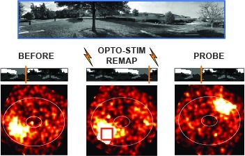
Many animals rely on an internal heading representation when navigating in varied environments. How this representation is linked to the sensory cues that define different surroundings is unclear. In the fly brain, heading is represented by 'compass' neurons that innervate a ring-shaped structure known as the ellipsoid body. Each compass neuron receives inputs from 'ring' neurons that are selective for particular visual features; this combination provides an ideal substrate for the extraction of directional information from a visual scene. Here we combine two-photon calcium imaging and optogenetics in tethered flying flies with circuit modelling, and show how the correlated activity of compass and visual neurons drives plasticity, which flexibly transforms two-dimensional visual cues into a stable heading representation. We also describe how this plasticity enables the fly to convert a partial heading representation, established from orienting within part of a novel setting, into a complete heading representation. Our results provide mechanistic insight into the memory-related computations that are essential for flexible navigation in varied surroundings.
Calcium imaging with genetically encoded calcium indicators (GECIs) is routinely used to measure neural activity in intact nervous systems. GECIs are frequently used in one of two different modes: to track activity in large populations of neuronal cell bodies, or to follow dynamics in subcellular compartments such as axons, dendrites and individual synaptic compartments. Despite major advances, calcium imaging is still limited by the biophysical properties of existing GECIs, including affinity, signal-to-noise ratio, rise and decay kinetics, and dynamic range. Using structure-guided mutagenesis and neuron-based screening, we optimized the green fluorescent protein-based GECI GCaMP6 for different modes of in vivo imaging. The jGCaMP7 sensors provide improved detection of individual spikes (jGCaMP7s,f), imaging in neurites and neuropil (jGCaMP7b), and tracking large populations of neurons using 2-photon (jGCaMP7s,f) or wide-field (jGCaMP7c) imaging.
The ability to measure synaptic connectivity and properties is essential for understanding neuronal circuits. However, existing methods that allow such measurements at cellular resolution are laborious and technically demanding. Here, we describe a system that allows such measurements in a high-throughput way by combining two-photon optogenetics and volumetric Ca2+ imaging with whole-cell recording. We reveal a circuit motif for generating fast undulatory locomotion in zebrafish.
Point-scanning two-photon microscopy enables high-resolution imaging within scattering specimens such as the mammalian brain, but sequential acquisition of voxels fundamentally limits imaging speed. We developed a two-photon imaging technique that scans lines of excitation across a focal plane at multiple angles and uses prior information to recover high-resolution images at over 1.4 billion voxels per second. Using a structural image as a prior for recording neural activity, we imaged visually-evoked and spontaneous glutamate release across hundreds of dendritic spines in mice at depths over 250 microns and frame-rates over 1 kHz. Dendritic glutamate transients in anaesthetized mice are synchronized within spatially-contiguous domains spanning tens of microns at frequencies ranging from 1-100 Hz. We demonstrate high-speed recording of acetylcholine and calcium sensors, 3D single-particle tracking, and imaging in densely-labeled cortex. Our method surpasses limits on the speed of raster-scanned imaging imposed by fluorescence lifetime.


