Filter
Associated Lab
Publication Date
7 Janelia Publications
Showing 1-7 of 7 resultsDrosophila brains contain numerous neurons that form complex circuits. These neurons are derived in stereotyped patterns from a fixed number of progenitors, called neuroblasts, and identifying individual neurons made by a neuroblast facilitates the reconstruction of neural circuits. An improved MARCM (mosaic analysis with a repressible cell marker) technique, called twin-spot MARCM, allows one to label the sister clones derived from a common progenitor simultaneously in different colors. It enables identification of every single neuron in an extended neuronal lineage based on the order of neuron birth. Here we report the first example, to our knowledge, of complete lineage analysis among neurons derived from a common neuroblast that relay olfactory information from the antennal lobe (AL) to higher brain centers. By identifying the sequentially derived neurons, we found that the neuroblast serially makes 40 types of AL projection neurons (PNs). During embryogenesis, one PN with multi-glomerular innervation and 18 uniglomerular PNs targeting 17 glomeruli of the adult AL are born. Many more PNs of 22 additional types, including four types of polyglomerular PNs, derive after the neuroblast resumes dividing in early larvae. Although different offspring are generated in a rather arbitrary sequence, the birth order strictly dictates the fate of each post-mitotic neuron, including the fate of programmed cell death. Notably, the embryonic progenitor has an altered temporal identity following each self-renewing asymmetric cell division. After larval hatching, the same progenitor produces multiple neurons for each cell type, but the number of neurons for each type is tightly regulated. These observations substantiate the origin-dependent specification of neuron types. Sequencing neuronal lineages will not only unravel how a complex brain develops but also permit systematic identification of neuron types for detailed structure and function analysis of the brain.
To study the complex synaptic interactions underpinning dendritic information processing in single neurons, experimenters require methods to mimic presynaptic neurotransmitter release at multiple sites with no physiological damage. We show that laser scanning systems built around large-aperture acousto-optic deflectors and high numerical aperture objective lenses provide the sub-millisecond, sub-micron precision necessary to achieve physiological, exogenous synaptic stimulation. Our laser scanning systems can produce the sophisticated spatio-temporal patterns of synaptic input that are necessary to investigate single-neuron dendritic physiology.
Mitochondria are difficult targets for microscopy because of their small size and highly compartmentalized, membranous interior. Super-resolution fluorescence microscopy methods have recently been developed that exceed the historical limits of optical imaging. Here we outline considerations and techniques in preparing to image the relative location of mitochondrial proteins using photoactivated localization microscopy (PALM). PALM and similar methods have the capacity to dramatically increase our ability to image proteins within mitochondria, and to expand our knowledge of the location of macromolecules beyond the current limits of immunoEM.
The neuropile of the Drosophila brain is subdivided into anatomically discrete compartments. Compartments are rich in terminal neurite branching and synapses; they are the neuropile domains in which signal processing takes place. Compartment boundaries are defined by more or less dense layers of glial cells as well as long neurite fascicles. These fascicles are formed during the larval period, when the approximately 100 neuronal lineages that constitute the Drosophila central brain differentiate. Each lineage forms an axon tract with a characteristic trajectory in the neuropile; groups of spatially related tracts congregate into the brain fascicles that can be followed from the larva throughout metamorphosis into the adult stage. Here we provide a map of the adult brain compartments and the relevant fascicles defining compartmental boundaries. We have identified the neuronal lineages contributing to each fascicle, which allowed us to compare compartments of the larval and adult brain directly. Most adult compartments can be recognized already in the early larval brain, where they form a "protomap" of the later adult compartments. Our analysis highlights the morphogenetic changes shaping the Drosophila brain; the data will be important for studies that link early-acting genetic mechanisms to the adult neuronal structures and circuits controlled by these mechanisms.
Recording light-microscopy images of large, nontransparent specimens, such as developing multicellular organisms, is complicated by decreased contrast resulting from light scattering. Early zebrafish development can be captured by standard light-sheet microscopy, but new imaging strategies are required to obtain high-quality data of late development or of less transparent organisms. We combined digital scanned laser light-sheet fluorescence microscopy with incoherent structured-illumination microscopy (DSLM-SI) and created structured-illumination patterns with continuously adjustable frequencies. Our method discriminates the specimen-related scattered background from signal fluorescence, thereby removing out-of-focus light and optimizing the contrast of in-focus structures. DSLM-SI provides rapid control of the illumination pattern, exceptional imaging quality, and high imaging speeds. We performed long-term imaging of zebrafish development for 58 h and fast multiple-view imaging of early Drosophila melanogaster development. We reconstructed cell positions over time from the Drosophila DSLM-SI data and created a fly digital embryo.
Although hippocampal theta oscillations represent a prime example of temporal coding in the mammalian brain, little is known about the specific biophysical mechanisms. Intracellular recordings support a particular abstract oscillatory interference model of hippocampal theta activity, the soma-dendrite interference model. To gain insight into the cellular and circuit level mechanisms of theta activity, we implemented a similar form of interference using the actual hippocampal network in mice in vitro. We found that pairing increasing levels of phasic dendritic excitation with phasic stimulation of perisomatic projecting inhibitory interneurons induced a somatic polarization and action potential timing profile that reproduced most common features. Alterations in the temporal profile of inhibition were required to fully capture all features. These data suggest that theta-related place cell activity is generated through an interaction between a phasic dendritic excitation and a phasic perisomatic shunting inhibition delivered by interneurons, a subset of which undergo activity-dependent presynaptic modulation.
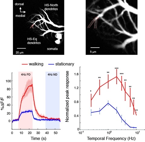
Changes in behavioral state modify neural activity in many systems. In some vertebrates such modulation has been observed and interpreted in the context of attention and sensorimotor coordinate transformations. Here we report state-dependent activity modulations during walking in a visual-motor pathway of Drosophila. We used two-photon imaging to monitor intracellular calcium activity in motion-sensitive lobula plate tangential cells (LPTCs) in head-fixed Drosophila walking on an air-supported ball. Cells of the horizontal system (HS)–a subgroup of LPTCs–showed stronger calcium transients in response to visual motion when flies were walking rather than resting. The amplified responses were also correlated with walking speed. Moreover, HS neurons showed a relatively higher gain in response strength at higher temporal frequencies, and their optimum temporal frequency was shifted toward higher motion speeds. Walking-dependent modulation of HS neurons in the Drosophila visual system may constitute a mechanism to facilitate processing of higher image speeds in behavioral contexts where these speeds of visual motion are relevant for course stabilization.