Filter
Associated Lab
- Ahrens Lab (2) Apply Ahrens Lab filter
- Aso Lab (1) Apply Aso Lab filter
- Baker Lab (2) Apply Baker Lab filter
- Betzig Lab (4) Apply Betzig Lab filter
- Bock Lab (2) Apply Bock Lab filter
- Cardona Lab (1) Apply Cardona Lab filter
- Cui Lab (2) Apply Cui Lab filter
- Dickson Lab (1) Apply Dickson Lab filter
- Druckmann Lab (1) Apply Druckmann Lab filter
- Dudman Lab (2) Apply Dudman Lab filter
- Eddy/Rivas Lab (2) Apply Eddy/Rivas Lab filter
- Egnor Lab (1) Apply Egnor Lab filter
- Fetter Lab (3) Apply Fetter Lab filter
- Gonen Lab (9) Apply Gonen Lab filter
- Grigorieff Lab (1) Apply Grigorieff Lab filter
- Harris Lab (3) Apply Harris Lab filter
- Heberlein Lab (1) Apply Heberlein Lab filter
- Hess Lab (2) Apply Hess Lab filter
- Jayaraman Lab (3) Apply Jayaraman Lab filter
- Ji Lab (1) Apply Ji Lab filter
- Karpova Lab (1) Apply Karpova Lab filter
- Keller Lab (9) Apply Keller Lab filter
- Lavis Lab (4) Apply Lavis Lab filter
- Leonardo Lab (3) Apply Leonardo Lab filter
- Looger Lab (10) Apply Looger Lab filter
- Magee Lab (3) Apply Magee Lab filter
- Menon Lab (3) Apply Menon Lab filter
- Reiser Lab (1) Apply Reiser Lab filter
- Riddiford Lab (5) Apply Riddiford Lab filter
- Rubin Lab (5) Apply Rubin Lab filter
- Scheffer Lab (3) Apply Scheffer Lab filter
- Schreiter Lab (5) Apply Schreiter Lab filter
- Spruston Lab (2) Apply Spruston Lab filter
- Stern Lab (5) Apply Stern Lab filter
- Sternson Lab (3) Apply Sternson Lab filter
- Svoboda Lab (10) Apply Svoboda Lab filter
- Tjian Lab (1) Apply Tjian Lab filter
- Truman Lab (3) Apply Truman Lab filter
- Wu Lab (3) Apply Wu Lab filter
- Zlatic Lab (2) Apply Zlatic Lab filter
Associated Project Team
Associated Support Team
Publication Date
- December 2013 (7) Apply December 2013 filter
- November 2013 (10) Apply November 2013 filter
- October 2013 (16) Apply October 2013 filter
- September 2013 (14) Apply September 2013 filter
- August 2013 (11) Apply August 2013 filter
- July 2013 (13) Apply July 2013 filter
- June 2013 (13) Apply June 2013 filter
- May 2013 (5) Apply May 2013 filter
- April 2013 (9) Apply April 2013 filter
- March 2013 (9) Apply March 2013 filter
- February 2013 (9) Apply February 2013 filter
- January 2013 (20) Apply January 2013 filter
- Remove 2013 filter 2013
136 Janelia Publications
Showing 41-50 of 136 resultsA central problem in neuroscience is reconstructing neuronal circuits on the synapse level. Due to a wide range of scales in brain architecture such reconstruction requires imaging that is both high-resolution and high-throughput. Existing electron microscopy (EM) techniques possess required resolution in the lateral plane and either high-throughput or high depth resolution but not both. Here, we exploit recent advances in unsupervised learning and signal processing to obtain high depth-resolution EM images computationally without sacrificing throughput. First, we show that the brain tissue can be represented as a sparse linear combination of localized basis functions that are learned using high-resolution datasets. We then develop compressive sensing-inspired techniques that can reconstruct the brain tissue from very few (typically 5) tomographic views of each section. This enables tracing of neuronal processes and, hence, high throughput reconstruction of neural circuits on the level of individual synapses.
Internal state as well as environmental conditions influence choice behavior. The neural circuits underpinning state-dependent behavior remain largely unknown. Carbon dioxide (CO2) is an important olfactory cue for many insects, including mosquitoes, flies, moths, and honeybees [1]. Concentrations of CO2 higher than 0.02% above atmospheric level trigger a strong innate avoidance in the fly Drosophila melanogaster [2, 3]. Here, we show that the mushroom body (MB), a brain center essential for olfactory associative memories [4-6] but thought to be dispensable for innate odor processing [7], is essential for CO2 avoidance behavior only in the context of starvation or in the context of a food-related odor. Consistent with this, CO2 stimulation elicits Ca(2+) influx into the MB intrinsic cells (Kenyon cells: KCs) in vivo. We identify an atypical projection neuron (bilateral ventral projection neuron, biVPN) that connects CO2 sensory input bilaterally to the MB calyx. Blocking synaptic output of the biVPN completely abolishes CO2 avoidance in food-deprived flies, but not in fed flies. These findings show that two alternative neural pathways control innate choice behavior, and they are dependent on the animal’s internal state. In addition, they suggest that, during innate choice behavior, the MB serves as an integration site for internal state and olfactory input.
An often-overlooked aspect of neural plasticity is the plasticity of neuronal composition, in which the numbers of neurons of particular classes are altered in response to environment and experience. The Drosophila brain features several well-characterized lineages in which a single neuroblast gives rise to multiple neuronal classes in a stereotyped sequence during development [1]. We find that in the intrinsic mushroom body neuron lineage, the numbers for each class are highly plastic, depending on the timing of temporal fate transitions and the rate of neuroblast proliferation. For example, mushroom body neuroblast cycling can continue under starvation conditions, uncoupled from temporal fate transitions that depend on extrinsic cues reflecting organismal growth and development. In contrast, the proliferation rates of antennal lobe lineages are closely associated with organismal development, and their temporal fate changes appear to be cell cycle-dependent, such that the same numbers and types of uniglomerular projection neurons innervate the antennal lobe following various perturbations. We propose that this surprising difference in plasticity for these brain lineages is adaptive, given their respective roles as parallel processors versus discrete carriers of olfactory information.

Optical flow is a key method used for quantitative motion estimation of biological structures in light microscopy. It has also been used as a key module in segmentation and tracking systems and is considered a mature technology in the field of computer vision. However, most of the research focused on 2D natural images, which are small in size and rich in edges and texture information. In contrast, 3D time-lapse recordings of biological specimens comprise up to several terabytes of image data and often exhibit complex object dynamics as well as blurring due to the point-spread-function of the microscope. Thus, new approaches to optical flow are required to improve performance for such data. We solve optical flow in large 3D time-lapse microscopy datasets by defining a Markov random field (MRF) over super-voxels in the foreground and applying motion smoothness constraints between super-voxels instead of voxel-wise. This model is tailored to the specific characteristics of light microscopy datasets: super-voxels help registration in textureless areas, the MRF over super-voxels efficiently propagates motion information between neighboring cells and the background subtraction and super-voxels reduce the dimensionality of the problem by an order of magnitude. We validate our approach on large 3D time-lapse datasets of Drosophila and zebrafish development by analyzing cell motion patterns. We show that our approach is, on average, 10 x faster than commonly used optical flow implementations in the Insight Tool-Kit (ITK) and reduces the average flow end point error by 50% in regions with complex dynamic processes, such as cell divisions.
Conventional acquisition of three-dimensional (3D) microscopy data requires sequential z scanning and is often too slow to capture biological events. We report an aberration-corrected multifocus microscopy method capable of producing an instant focal stack of nine 2D images. Appended to an epifluorescence microscope, the multifocus system enables high-resolution 3D imaging in multiple colors with single-molecule sensitivity, at speeds limited by the camera readout time of a single image.
Gap junctions (GJs) represent connexin-rich membrane domains that connect interiors of adjoining cells in mammalian tissues. How fast GJs can respond to bacterial pathogens has not been known previously. Using Bessel beam plane illumination and confocal spinning disk microscopy, we found fast ( 500 ms) formation of connexin-depleted regions (CDRs) inside GJ plaques between cells exposed to AB5 toxins. CDR formation appears as a fast redistribution of connexin channels within GJ plaques with minor changes in outline or geometry. CDR formation does not depend on membrane trafficking or submembrane cytoskeleton and has no effect on GJ conductance. However, CDR responses depend on membrane lipids, can be modified by cholesterol-clustering agents and extracellular K(+) ion concentration, and influence cAMP signaling. The CDR response of GJ plaques to bacterial toxins is a phenomenon observed for all tested connexin isoforms. Through signaling, the CDR response may enable cells to sense exposure to AB5 toxins. CDR formation may reflect lipid-phase separation events in the biological membrane of the GJ plaque, leading to increased connexin packing and lipid reorganization. Our data demonstrate very fast dynamics (in the millisecond-to-second range) within GJ plaques, which previously were considered to be relatively stable, long-lived structures.
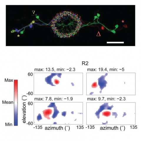
Many animals, including insects, are known to use visual landmarks to orient in their environment. In Drosophila melanogaster, behavioural genetics studies have identified a higher brain structure called the central complex as being required for the fly’s innate responses to vertical visual features and its short- and long-term memory for visual patterns. But whether and how neurons of the fly central complex represent visual features are unknown. Here we use two-photon calcium imaging in head-fixed walking and flying flies to probe visuomotor responses of ring neurons—a class of central complex neurons that have been implicated in landmark-driven spatial memory in walking flies and memory for visual patterns in tethered flying flies. We show that dendrites of ring neurons are visually responsive and arranged retinotopically. Ring neuron receptive fields comprise both excitatory and inhibitory subfields, resembling those of simple cells in the mammalian primary visual cortex. Ring neurons show strong and, in some cases, direction-selective orientation tuning, with a notable preference for vertically oriented features similar to those that evoke innate responses in flies. Visual responses were diminished during flight, but, in contrast with the hypothesized role of the central complex in the control of locomotion, not modulated during walking. Taken together, these results indicate that ring neurons represent behaviourally relevant visual features in the fly’s environment, enabling downstream central complex circuits to produce appropriate motor commands. More broadly, this study opens the door to mechanistic investigations of circuit computations underlying visually guided action selection in the Drosophila central complex.
In vertebrates, primary sex determination refers to the decision within a bipotential organ precursor to differentiate as a testis or ovary. Bifurcation of organ fate begins between embryonic day (E) 11.0–E12.0 in mice and likely involves a dynamic transcription network that is poorly understood. To elucidate the first steps of sexual fate specification, we profiled the XX and XY gonad transcriptomes at fine granularity during this period and resolved cascades of gene activation and repression. C57BL/6J (B6) XY gonads showed a consistent 5-hour delay in the activation of most male pathway genes and repression of female pathway genes relative to 129S1/SvImJ, which likely explains the sensitivity of the B6 strain to male-to-female sex reversal. Using this fine time course data, we predicted novel regulatory genes underlying expression QTLs (eQTLs) mapped in a previous study. To test predictions, we developed an in vitro gonad primary cell assay and optimized a lentivirus-based shRNA delivery method to silence candidate genes and quantify effects on putative targets. We provide strong evidence that Lmo4 (Lim-domain only 4) is a novel regulator of sex determination upstream of SF1 (Nr5a1), Sox9, Fgf9, and Col9a3. This approach can be readily applied to identify regulatory interactions in other systems.
Genetically hard-wired neural mechanisms must enforce behavioral reproductive isolation because interspecies courtship is rare even in sexually na{\"ıve animals of most species. We find that the chemoreceptor Gr32a inhibits male D. melanogaster from courting diverse fruit fly species. Gr32a recognizes nonvolatile aversive cues present on these reproductively dead-end targets, and activity of Gr32a neurons is necessary and sufficient to inhibit interspecies courtship. Male-specific Fruitless (Fru(M)), a master regulator of courtship, also inhibits interspecies courtship. Gr32a and Fru(M) are not coexpressed, but Fru(M) neurons contact Gr32a neurons, suggesting that these genes influence a shared neural circuit that inhibits interspecies courtship. Gr32a and Fru(M) also suppress within-species intermale courtship, but we show that distinct mechanisms preclude sexual displays toward conspecific males and other species. Although this chemosensory pathway does not inhibit interspecies mating in D. melanogaster females, similar mechanisms appear to inhibit this behavior in many other male drosophilids.
The discovery of intracellular Ca(2+) signals within astrocytes has changed our view of how these ubiquitous cells contribute to brain function. Classically thought merely to serve supportive functions, astrocytes are increasingly thought to respond to, and regulate, neurons. The use of organic Ca(2+) indicator dyes such as Fluo-4 and Fura-2 has proved instrumental in the study of astrocyte physiology. However, progress has recently been accelerated by the use of cytosolic and membrane targeted genetically encoded calcium indicators (GECIs). Herein, we review these recent findings, discuss why studying astrocyte Ca(2+) signals is important to understand brain function, and summarize work that led to the discovery of TRPA1 channel-mediated near-membrane Ca(2+) signals in astrocytes and their indirect neuromodulatory roles at inhibitory synapses in the CA1 stratum radiatum region of the hippocampus. We suggest that the use of membrane-targeted and cytosolic GECIs holds great promise to explore the diversity of Ca(2+) signals within single astrocytes and also to study diversity of function for astrocytes in different parts of the brain.