Filter
Associated Lab
- Aguilera Castrejon Lab (4) Apply Aguilera Castrejon Lab filter
- Ahrens Lab (64) Apply Ahrens Lab filter
- Aso Lab (42) Apply Aso Lab filter
- Baker Lab (19) Apply Baker Lab filter
- Betzig Lab (104) Apply Betzig Lab filter
- Beyene Lab (10) Apply Beyene Lab filter
- Bock Lab (14) Apply Bock Lab filter
- Branson Lab (53) Apply Branson Lab filter
- Card Lab (37) Apply Card Lab filter
- Cardona Lab (45) Apply Cardona Lab filter
- Chklovskii Lab (10) Apply Chklovskii Lab filter
- Clapham Lab (15) Apply Clapham Lab filter
- Cui Lab (19) Apply Cui Lab filter
- Darshan Lab (8) Apply Darshan Lab filter
- Dennis Lab (2) Apply Dennis Lab filter
- Dickson Lab (32) Apply Dickson Lab filter
- Druckmann Lab (21) Apply Druckmann Lab filter
- Dudman Lab (46) Apply Dudman Lab filter
- Eddy/Rivas Lab (30) Apply Eddy/Rivas Lab filter
- Egnor Lab (4) Apply Egnor Lab filter
- Espinosa Medina Lab (21) Apply Espinosa Medina Lab filter
- Feliciano Lab (16) Apply Feliciano Lab filter
- Fetter Lab (31) Apply Fetter Lab filter
- FIB-SEM Technology (1) Apply FIB-SEM Technology filter
- Fitzgerald Lab (17) Apply Fitzgerald Lab filter
- Freeman Lab (15) Apply Freeman Lab filter
- Funke Lab (46) Apply Funke Lab filter
- Gonen Lab (59) Apply Gonen Lab filter
- Grigorieff Lab (34) Apply Grigorieff Lab filter
- Harris Lab (55) Apply Harris Lab filter
- Heberlein Lab (13) Apply Heberlein Lab filter
- Hermundstad Lab (28) Apply Hermundstad Lab filter
- Hess Lab (77) Apply Hess Lab filter
- Ilanges Lab (4) Apply Ilanges Lab filter
- Jayaraman Lab (45) Apply Jayaraman Lab filter
- Ji Lab (33) Apply Ji Lab filter
- Johnson Lab (2) Apply Johnson Lab filter
- Karpova Lab (14) Apply Karpova Lab filter
- Keleman Lab (8) Apply Keleman Lab filter
- Keller Lab (62) Apply Keller Lab filter
- Koay Lab (4) Apply Koay Lab filter
- Lavis Lab (150) Apply Lavis Lab filter
- Lee (Albert) Lab (29) Apply Lee (Albert) Lab filter
- Leonardo Lab (19) Apply Leonardo Lab filter
- Li Lab (8) Apply Li Lab filter
- Lippincott-Schwartz Lab (110) Apply Lippincott-Schwartz Lab filter
- Liu (Yin) Lab (4) Apply Liu (Yin) Lab filter
- Liu (Zhe) Lab (60) Apply Liu (Zhe) Lab filter
- Looger Lab (137) Apply Looger Lab filter
- Magee Lab (31) Apply Magee Lab filter
- Menon Lab (12) Apply Menon Lab filter
- Murphy Lab (6) Apply Murphy Lab filter
- O'Shea Lab (7) Apply O'Shea Lab filter
- Otopalik Lab (1) Apply Otopalik Lab filter
- Pachitariu Lab (44) Apply Pachitariu Lab filter
- Pastalkova Lab (6) Apply Pastalkova Lab filter
- Pavlopoulos Lab (7) Apply Pavlopoulos Lab filter
- Pedram Lab (4) Apply Pedram Lab filter
- Podgorski Lab (16) Apply Podgorski Lab filter
- Reiser Lab (49) Apply Reiser Lab filter
- Riddiford Lab (20) Apply Riddiford Lab filter
- Romani Lab (40) Apply Romani Lab filter
- Rubin Lab (111) Apply Rubin Lab filter
- Saalfeld Lab (49) Apply Saalfeld Lab filter
- Satou Lab (3) Apply Satou Lab filter
- Scheffer Lab (38) Apply Scheffer Lab filter
- Schreiter Lab (55) Apply Schreiter Lab filter
- Schulze Lab (1) Apply Schulze Lab filter
- Sgro Lab (3) Apply Sgro Lab filter
- Shroff Lab (35) Apply Shroff Lab filter
- Simpson Lab (18) Apply Simpson Lab filter
- Singer Lab (37) Apply Singer Lab filter
- Spruston Lab (62) Apply Spruston Lab filter
- Stern Lab (77) Apply Stern Lab filter
- Sternson Lab (47) Apply Sternson Lab filter
- Stringer Lab (41) Apply Stringer Lab filter
- Svoboda Lab (132) Apply Svoboda Lab filter
- Tebo Lab (12) Apply Tebo Lab filter
- Tervo Lab (10) Apply Tervo Lab filter
- Tillberg Lab (19) Apply Tillberg Lab filter
- Tjian Lab (17) Apply Tjian Lab filter
- Truman Lab (58) Apply Truman Lab filter
- Turaga Lab (41) Apply Turaga Lab filter
- Turner Lab (27) Apply Turner Lab filter
- Vale Lab (8) Apply Vale Lab filter
- Voigts Lab (5) Apply Voigts Lab filter
- Wang (Meng) Lab (31) Apply Wang (Meng) Lab filter
- Wang (Shaohe) Lab (6) Apply Wang (Shaohe) Lab filter
- Wong-Campos Lab (1) Apply Wong-Campos Lab filter
- Wu Lab (8) Apply Wu Lab filter
- Zlatic Lab (26) Apply Zlatic Lab filter
- Zuker Lab (5) Apply Zuker Lab filter
Associated Project Team
- CellMap (13) Apply CellMap filter
- COSEM (3) Apply COSEM filter
- FIB-SEM Technology (5) Apply FIB-SEM Technology filter
- Fly Descending Interneuron (12) Apply Fly Descending Interneuron filter
- Fly Functional Connectome (14) Apply Fly Functional Connectome filter
- Fly Olympiad (5) Apply Fly Olympiad filter
- FlyEM (56) Apply FlyEM filter
- FlyLight (50) Apply FlyLight filter
- GENIE (47) Apply GENIE filter
- Integrative Imaging (11) Apply Integrative Imaging filter
- Larval Olympiad (2) Apply Larval Olympiad filter
- MouseLight (18) Apply MouseLight filter
- NeuroSeq (1) Apply NeuroSeq filter
- ThalamoSeq (1) Apply ThalamoSeq filter
- Tool Translation Team (T3) (29) Apply Tool Translation Team (T3) filter
- Transcription Imaging (45) Apply Transcription Imaging filter
Associated Support Team
- Project Pipeline Support (5) Apply Project Pipeline Support filter
- Anatomy and Histology (18) Apply Anatomy and Histology filter
- Cryo-Electron Microscopy (49) Apply Cryo-Electron Microscopy filter
- Electron Microscopy (18) Apply Electron Microscopy filter
- Gene Targeting and Transgenics (11) Apply Gene Targeting and Transgenics filter
- High Performance Computing (7) Apply High Performance Computing filter
- Integrative Imaging (25) Apply Integrative Imaging filter
- Invertebrate Shared Resource (40) Apply Invertebrate Shared Resource filter
- Janelia Experimental Technology (40) Apply Janelia Experimental Technology filter
- Management Team (1) Apply Management Team filter
- Mass Spectrometry (1) Apply Mass Spectrometry filter
- Molecular Genomics (15) Apply Molecular Genomics filter
- Project Technical Resources (54) Apply Project Technical Resources filter
- Quantitative Genomics (20) Apply Quantitative Genomics filter
- Scientific Computing (106) Apply Scientific Computing filter
- Stem Cell & Primary Culture (14) Apply Stem Cell & Primary Culture filter
- Viral Tools (14) Apply Viral Tools filter
- Vivarium (7) Apply Vivarium filter
Publication Date
- 2026 (118) Apply 2026 filter
- 2025 (223) Apply 2025 filter
- 2024 (208) Apply 2024 filter
- 2023 (157) Apply 2023 filter
- 2022 (166) Apply 2022 filter
- 2021 (175) Apply 2021 filter
- 2020 (177) Apply 2020 filter
- 2019 (177) Apply 2019 filter
- 2018 (206) Apply 2018 filter
- 2017 (186) Apply 2017 filter
- 2016 (191) Apply 2016 filter
- 2015 (195) Apply 2015 filter
- 2014 (190) Apply 2014 filter
- 2013 (136) Apply 2013 filter
- 2012 (112) Apply 2012 filter
- 2011 (98) Apply 2011 filter
- 2010 (61) Apply 2010 filter
- 2009 (56) Apply 2009 filter
- 2008 (40) Apply 2008 filter
- 2007 (21) Apply 2007 filter
- 2006 (3) Apply 2006 filter
2896 Janelia Publications
Showing 1761-1770 of 2896 results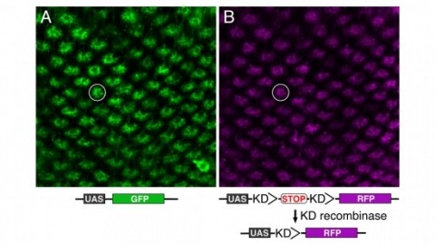
Site-specific recombinases have been used for two decades to manipulate the structure of animal genomes in highly predictable ways and have become major research tools. However, the small number of recombinases demonstrated to have distinct specificities, low toxicity, and sufficient activity to drive reactions to completion in animals has been a limitation. In this report we show that four recombinases derived from yeast-KD, B2, B3, and R-are highly active and nontoxic in Drosophila and that KD, B2, B3, and the widely used FLP recombinase have distinct target specificities. We also show that the KD and B3 recombinases are active in mice.
Glomeruli are functional units in the olfactory system. The mouse olfactory bulb contains roughly 2,000 glomeruli, each receiving inputs from olfactory sensory neurons (OSNs) that express a specific odorant receptor gene. Odors typically activate many glomeruli in complex combinatorial patterns and it is unknown which features of neuronal activity in individual glomeruli contribute to odor perception. To address this, we used optogenetics to selectively activate single, genetically identified glomeruli in behaving mice. We found that mice could perceive the stimulation of a single glomerulus. Single-glomerulus stimulation was also detected on an intense odor background. In addition, different input intensities and the timing of input relative to sniffing were discriminated through one glomerulus. Our data suggest that each glomerulus can transmit odor information using identity, intensity and temporal coding cues. These multiple modes of information transmission may enable the olfactory system to efficiently identify and localize odor sources.
Understanding gene expression is essential for deciphering cellular functions. However, methods for analyzing the expression of numerous genes in situ within a given tissue remain limited. The EASI-FISH protocol described here has been adapted to detect the expression of dozens of genes in the intact adult Drosophila central nervous system (CNS) using commercially available reagents. This protocol includes a new gel formulation that enhances gel robustness, enabling multiple rounds of hybridization and allowing the embedding of multiple brains per gel. This improvement increases throughput, facilitates optimal comparison of experimental conditions, and reduces reagent costs. Additionally, by employing the GAL4-UAS system for co-detection of green fluorescent protein (GFP), gene expression can be visualized in specific neuronal or glial cell types. Notably, the high resolution achieved through expansion microscopy, combined with the sensitivity of the method, allows for the detection of single RNA transcripts. This approach effectively integrates high image quality with high throughput, making it a powerful tool for studying gene expression throughout the intact fly brain.
The lack of efficient tools to label multiple endogenous targets in cell lines without staining or fixation has limited our ability to track physiological and pathological changes in cells over time via live-cell studies. Here, we outline the FAST-HDR vector system to be used in combination with CRISPR-Cas9 to allow visual live-cell studies of up to three endogenous proteins within the same cell line. Our approach utilizes a novel set of advanced donor plasmids for homology-directed repair and a streamlined workflow optimized for microscopy-based cell screening to create genetically modified cell lines that do not require staining or fixation to accommodate microscopy-based studies. We validated this new methodology by developing two advanced cell lines with three fluorescent-labeled endogenous proteins that support high-content imaging without using antibodies or exogenous staining. We applied this technology to study seven severe acute respiratory syndrome coronavirus 2 (SARS-CoV-2/COVID-19) viral proteins to understand better their effects on autophagy, mitochondrial dynamics, and cell growth. Using these two cell lines, we were able to identify the protein ORF3a successfully as a potent inhibitor of autophagy, inducer of mitochondrial relocalization, and a growth inhibitor, which highlights the effectiveness of live-cell studies using this technology.
Information processing by brain circuits depends on Ca-dependent, stochastic release of the excitatory neurotransmitter glutamate. Whilst optical glutamate sensors have enabled detection of synaptic discharges, understanding presynaptic machinery requires simultaneous readout of glutamate release and nanomolar presynaptic Ca in situ. Here, we find that the fluorescence lifetime of the red-shifted Ca indicator Cal-590 is Ca-sensitive in the nanomolar range, and employ it in combination with green glutamate sensors to relate quantal neurotransmission to presynaptic Ca kinetics. Multiplexed imaging of individual and multiple synapses in identified axonal circuits reveals that glutamate release efficacy, but not its short-term plasticity, varies with time-dependent fluctuations in presynaptic resting Ca or spike-evoked Ca entry. Within individual presynaptic boutons, we find no nanoscopic co-localisation of evoked presynaptic Ca entry with the prevalent glutamate release site, suggesting loose coupling between the two. The approach enables a better understanding of release machinery at central synapses.
3-photon excitation enables in vivo fluorescence microscopy deep in densely labeled and highly scattering samples, while maintaining high resolution and contrast. We designed and characterized a dual-plane 3-photon microscope with temporal multiplexing and remote focusing, and performed simultaneous in vivo calcium imaging of two planes deep in the cortex of a transgenic mouse expressing GCaMP6s in nearly all excitatory neurons.
We describe an adaptive optics method that modulates the intensity or phase of light rays at multiple pupil segments in parallel to determine the sample-induced aberration. Applicable to fluorescent protein-labeled structures of arbitrary complexity, it allowed us to obtain diffraction-limited resolution in various samples in vivo. For the strongly scattering mouse brain, a single aberration correction improved structural and functional imaging of fine neuronal processes over a large imaging volume.
We consider the problem of optimizing general convex objective functions with nonnegativity constraints. Using the Karush-Kuhn-Tucker (KKT) conditions for the nonnegativity constraints we will derive fast multiplicative update rules for several problems of interest in signal processing, including non-negative deconvolution, point-process smoothing, ML estimation for Poisson Observations, nonnegative least squares and nonnegative matrix factorization (NMF). Our algorithm can also account for temporal and spatial structure and regularization. We will analyze the performance of our algorithm on simultaneously recorded neuronal calcium imaging and electrophysiology data.
Methods of three-dimensional electron microscopy have been actively developed recently and open up great opportunities for morphological work. This approach is especially useful for studying microinsects, since it is possible to obtain complete series of high-resolution sections of a whole insect. Studies on the genus Megaphragma are especially important, since the unique phenomenon of lysis of most of the neuron nuclei was discovered in species of this genus. In this study we reveal the anatomical structure of the head of Megaphragma viggianii at all levels from organs to subcellular structures. Despite the miniature size of the body, most of the organ systems of M. viggianii retain the structural plan and complexity of organization at all levels. The set of muscles and the well-developed stomatogastric nervous system of this species correspond to those of larger insects, and there is also a well-developed tracheal system in the head of this species. Reconstructions of the head of M. viggianii at the cellular and subcellular levels were obtained, and of volumetric data were analyzed. A total of 689 nucleated cells of the head were reconstructed. The ultrastructure of M. viggianii is surprisingly complex, and the evolutionary benefits of such complexity are probably among the factors limiting the further miniaturization of parasitoid wasps.
The structure and interaction networks of molecules within biomolecular condensates are poorly understood. Using cryo-electron tomography and molecular dynamics simulations, we elucidated the structure of phase-separated chromatin condensates across scales, from individual amino acids to network architecture. We found that internucleosomal DNA linker length controls nucleosome arrangement and histone tail interactions, shaping the structure of individual chromatin molecules within and outside condensates. This structural modulation determines the balance between intra- and intermolecular interactions, which governs the molecular network, thermodynamic stability, and material properties of chromatin condensates. Mammalian nuclei contain dense clusters of nucleosomes whose nonrandom organization is mirrored by the reconstituted condensates. Our work explains how the structure of individual chromatin molecules determines physical properties of chromatin condensates and cellular chromatin organization.