Filter
Associated Lab
- Ahrens Lab (2) Apply Ahrens Lab filter
- Aso Lab (3) Apply Aso Lab filter
- Baker Lab (1) Apply Baker Lab filter
- Betzig Lab (8) Apply Betzig Lab filter
- Bock Lab (1) Apply Bock Lab filter
- Branson Lab (7) Apply Branson Lab filter
- Card Lab (4) Apply Card Lab filter
- Cardona Lab (8) Apply Cardona Lab filter
- Cui Lab (1) Apply Cui Lab filter
- Dickson Lab (1) Apply Dickson Lab filter
- Druckmann Lab (3) Apply Druckmann Lab filter
- Dudman Lab (4) Apply Dudman Lab filter
- Eddy/Rivas Lab (1) Apply Eddy/Rivas Lab filter
- Feliciano Lab (1) Apply Feliciano Lab filter
- Fetter Lab (4) Apply Fetter Lab filter
- Funke Lab (1) Apply Funke Lab filter
- Gonen Lab (11) Apply Gonen Lab filter
- Grigorieff Lab (6) Apply Grigorieff Lab filter
- Harris Lab (5) Apply Harris Lab filter
- Heberlein Lab (1) Apply Heberlein Lab filter
- Hermundstad Lab (1) Apply Hermundstad Lab filter
- Hess Lab (4) Apply Hess Lab filter
- Jayaraman Lab (4) Apply Jayaraman Lab filter
- Ji Lab (5) Apply Ji Lab filter
- Keleman Lab (1) Apply Keleman Lab filter
- Keller Lab (2) Apply Keller Lab filter
- Lavis Lab (16) Apply Lavis Lab filter
- Lee (Albert) Lab (6) Apply Lee (Albert) Lab filter
- Leonardo Lab (2) Apply Leonardo Lab filter
- Lippincott-Schwartz Lab (9) Apply Lippincott-Schwartz Lab filter
- Liu (Zhe) Lab (5) Apply Liu (Zhe) Lab filter
- Looger Lab (6) Apply Looger Lab filter
- Magee Lab (2) Apply Magee Lab filter
- Menon Lab (1) Apply Menon Lab filter
- Pachitariu Lab (1) Apply Pachitariu Lab filter
- Reiser Lab (6) Apply Reiser Lab filter
- Riddiford Lab (1) Apply Riddiford Lab filter
- Romani Lab (6) Apply Romani Lab filter
- Rubin Lab (15) Apply Rubin Lab filter
- Saalfeld Lab (4) Apply Saalfeld Lab filter
- Scheffer Lab (4) Apply Scheffer Lab filter
- Schreiter Lab (4) Apply Schreiter Lab filter
- Shroff Lab (1) Apply Shroff Lab filter
- Simpson Lab (2) Apply Simpson Lab filter
- Singer Lab (6) Apply Singer Lab filter
- Spruston Lab (1) Apply Spruston Lab filter
- Stern Lab (8) Apply Stern Lab filter
- Sternson Lab (2) Apply Sternson Lab filter
- Svoboda Lab (9) Apply Svoboda Lab filter
- Truman Lab (6) Apply Truman Lab filter
- Turaga Lab (3) Apply Turaga Lab filter
- Turner Lab (2) Apply Turner Lab filter
- Wu Lab (1) Apply Wu Lab filter
- Zlatic Lab (7) Apply Zlatic Lab filter
Associated Project Team
- Fly Descending Interneuron (1) Apply Fly Descending Interneuron filter
- Fly Functional Connectome (4) Apply Fly Functional Connectome filter
- Fly Olympiad (1) Apply Fly Olympiad filter
- FlyEM (4) Apply FlyEM filter
- FlyLight (2) Apply FlyLight filter
- GENIE (3) Apply GENIE filter
- ThalamoSeq (1) Apply ThalamoSeq filter
- Tool Translation Team (T3) (3) Apply Tool Translation Team (T3) filter
- Transcription Imaging (6) Apply Transcription Imaging filter
Associated Support Team
- Anatomy and Histology (2) Apply Anatomy and Histology filter
- Cryo-Electron Microscopy (4) Apply Cryo-Electron Microscopy filter
- Electron Microscopy (1) Apply Electron Microscopy filter
- Integrative Imaging (1) Apply Integrative Imaging filter
- Invertebrate Shared Resource (1) Apply Invertebrate Shared Resource filter
- Project Technical Resources (1) Apply Project Technical Resources filter
- Quantitative Genomics (2) Apply Quantitative Genomics filter
- Scientific Computing Software (9) Apply Scientific Computing Software filter
- Viral Tools (1) Apply Viral Tools filter
- Vivarium (1) Apply Vivarium filter
Publication Date
- December 2017 (15) Apply December 2017 filter
- November 2017 (11) Apply November 2017 filter
- October 2017 (7) Apply October 2017 filter
- September 2017 (14) Apply September 2017 filter
- August 2017 (15) Apply August 2017 filter
- July 2017 (20) Apply July 2017 filter
- June 2017 (18) Apply June 2017 filter
- May 2017 (25) Apply May 2017 filter
- April 2017 (21) Apply April 2017 filter
- March 2017 (15) Apply March 2017 filter
- February 2017 (7) Apply February 2017 filter
- January 2017 (18) Apply January 2017 filter
- Remove 2017 filter 2017
186 Janelia Publications
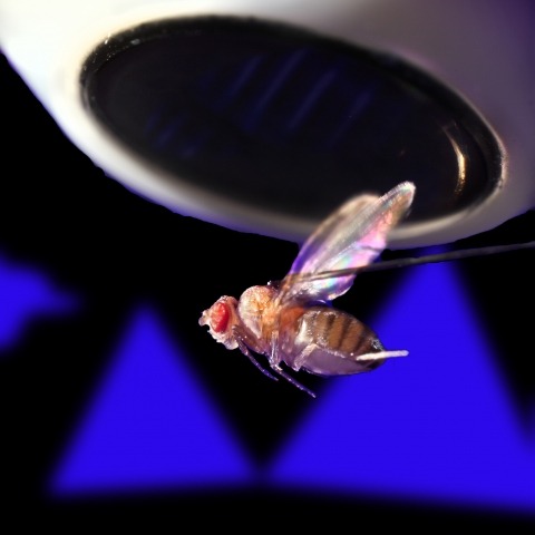
Showing 121-130 of 186 resultsNervous systems have evolved to translate external stimuli into appropriate behavioral responses. In an ever-changing environment, flexible adjustment of behavioral choice by experience-dependent learning is essential for the animal's survival. Associative learning is a simple form of learning that is widely observed from worms to humans. To understand the whole process of learning, we need to know how sensory information is represented and transformed in the brain, how it is changed by experience, and how the changes are reflected on motor output. To tackle these questions, studying numerically simple invertebrate nervous systems has a great advantage. In this review, I will feature the Pavlovian olfactory learning in the fruit fly, Drosophila melanogaster. The mushroom body is a key brain area for the olfactory learning in this organism. Recently, comprehensive anatomical information and the genetic tool sets were made available for the mushroom body circuit. This greatly accelerated the physiological understanding of the learning process. One of the key findings was dopamine-induced long-term synaptic plasticity that can alter the representations of stimulus valence. I will mostly focus on the new studies within these few years and discuss what we can possibly learn about the vertebrate systems from this model organism.
Ring attractors are a class of recurrent networks hypothesized to underlie the representation of heading direction. Such network structures, schematized as a ring of neurons whose connectivity depends on their heading preferences, can sustain a bump-like activity pattern whose location can be updated by continuous shifts along either turn direction. We recently reported that a population of fly neurons represents the animal's heading via bump-like activity dynamics. We combined two-photon calcium imaging in head-fixed flying flies with optogenetics to overwrite the existing population representation with an artificial one, which was then maintained by the circuit with naturalistic dynamics. A network with local excitation and global inhibition enforces this unique and persistent heading representation. Ring attractor networks have long been invoked in theoretical work; our study provides physiological evidence of their existence and functional architecture.
Persistent neural activity maintains information that connects past and future events. Models of persistent activity often invoke reverberations within local cortical circuits, but long-range circuits could also contribute. Neurons in the mouse anterior lateral motor cortex (ALM) have been shown to have selective persistent activity that instructs future actions. The ALM is connected bidirectionally with parts of the thalamus, including the ventral medial and ventral anterior-lateral nuclei. We recorded spikes from the ALM and thalamus during tactile discrimination with a delayed directional response. Here we show that, similar to ALM neurons, thalamic neurons exhibited selective persistent delay activity that predicted movement direction. Unilateral photoinhibition of delay activity in the ALM or thalamus produced contralesional neglect. Photoinhibition of the thalamus caused a short-latency and near-complete collapse of ALM activity. Similarly, photoinhibition of the ALM diminished thalamic activity. Our results show that the thalamus is a circuit hub in motor preparation and suggest that persistent activity requires reciprocal excitation across multiple brain areas.
We present an approach to study macromolecular assemblies by detecting component proteins' characteristic high-resolution projection patterns, calculated from their known 3D structures, in single electron cryo-micrographs. Our method detects single apoferritin molecules in vitreous ice with high specificity and determines their orientation and location precisely. Simulations show that high spatial-frequency information and-in the presence of protein background-a whitening filter are essential for optimal detection, in particular for images taken far from focus. Experimentally, we could detect small viral RNA polymerase molecules, distributed randomly among binding locations, inside rotavirus particles. Based on the currently attainable image quality, we estimate a threshold for detection that is 150 kDa in ice and 300 kDa in 100 nm thick samples of dense biological material.
Animal development is orchestrated by spatio-temporal gene expression programmes that drive precise lineage commitment, proliferation and migration events at the single-cell level, collectively leading to large-scale morphological change and functional specification in the whole organism. Efforts over decades have uncovered two 'seemingly contradictory' mechanisms in gene regulation governing these intricate processes: (i) stochasticity at individual gene regulatory steps in single cells and (ii) highly coordinated gene expression dynamics in the embryo. Here we discuss how these two layers of regulation arise from the molecular and the systems level, and how they might interplay to determine cell fate and to control the complex body plan. We also review recent technological advancements that enable quantitative analysis of gene regulation dynamics at single-cell, single-molecule resolution. These approaches outline next-generation experiments to decipher general principles bridging gaps between molecular dynamics in single cells and robust gene regulations in the embryo.
Insect nervous systems are proven and powerful model systems for neuroscience research with wide relevance in biology and medicine. However, descriptions of insect brains have suffered from a lack of a complete and uniform nomenclature. Recognising this problem the Insect Brain Name Working Group produced the first agreed hierarchical nomenclature system for the adult insect brain, using Drosophila melanogaster as the reference framework, with other insect taxa considered to ensure greater consistency and expandability (Ito et al., 2014). Ito et al. (2014) purposely focused on the gnathal regions that account for approximately 50% of the adult CNS. We extend this nomenclature system to the sub-gnathal regions of the adult Drosophila nervous system to provide a nomenclature of the so-called ventral nervous system (VNS), which includes the thoracic and abdominal neuromeres that was not included in the original work and contains the neurons that play critical roles underpinning most fly behaviours.
Mycobacteria possess a multi-layered cell wall that requires extensive remodelling during cell division. We investigated the role of an amidase_3 domain-containing N-acetylmuramyl-L-alanine amidase, a peptidoglycan remodelling enzyme implicated in cell division. We demonstrated that deletion of MSMEG_6281 (Ami1) in Mycobacterium smegmatis resulted in the formation of cellular chains, illustrative of cells that were unable to complete division. Suprisingly, viability in the Δami1 mutant was maintained through atypical lateral branching, the products of which proceeded to form viable daughter cells. We showed that these lateral buds resulted from mislocalization of DivIVA, a major determinant in facilitating polar elongation in mycobacterial cells. Failure of Δami1 mutant cells to separate also led to dysregulation of FtsZ ring bundling. Loss of Ami1 resulted in defects in septal peptidoglycan turnover with release of excess cell wall material from the septum or newly born cell poles. We noted signficant accumulation of 3-3 crosslinked muropeptides in the Δami1 mutant. We further demonstrated that deletion of ami1 leads to increased cell wall permeability and enhanced susceptiblity to cell wall targeting antibiotics. Collectively, these data provide novel insight on cell division in actinobacteria and highlights a new class of potential drug targets for mycobacterial diseases.
The cortical actin cytoskeleton has been shown to be critical for the reorganization and heterogeneity of plasma membrane components of many cells, including T cells. Building on previous studies at the T cell immunological synapse, we quantitatively assess the structure and dynamics of this meshwork using live-cell superresolution fluorescence microscopy and spatio-temporal image correlation spectroscopy. We show for the first time, to our knowledge, that not only does the dense actin cortex flow in a retrograde fashion toward the synapse center, but the plasma membrane itself shows similar behavior. Furthermore, using two-color, live-cell superresolution cross-correlation spectroscopy, we demonstrate that the two flows are correlated and, in addition, we show that coupling may extend to the outer leaflet of the plasma membrane by examining the flow of GPI-anchored proteins. Finally, we demonstrate that the actin flow is correlated with a third component, α-actinin, which upon CRISPR knockout led to reduced plasma membrane flow directionality despite increased actin flow velocity. We hypothesize that this apparent cytoskeletal-membrane coupling could provide a mechanism for driving the observed retrograde flow of signaling molecules such as the TCR, Lck, ZAP70, LAT, and SLP76.
Analysing computations in neural circuits often uses simplified models because the actual neuronal implementation is not known. For example, a problem in vision, how the eye detects image motion, has long been analysed using Hassenstein-Reichardt (HR) detector or Barlow-Levick (BL) models. These both simulate motion detection well, but the exact neuronal circuits undertaking these tasks remain elusive. We reconstructed a comprehensive connectome of the circuits of Drosophila's motion-sensing T4 cells using a novel EM technique. We uncover complex T4 inputs and reveal that putative excitatory inputs cluster at T4's dendrite shafts, while inhibitory inputs localize to the bases. Consistent with our previous study, we reveal that Mi1 and Tm3 cells provide most synaptic contacts onto T4. We are, however, unable to reproduce the spatial offset between these cells reported previously. Our comprehensive connectome reveals complex circuits that include candidate anatomical substrates for both HR and BL types of motion detectors.
Larval Drosophila offer a study case for behavioral neurogenetics that is simple enough to be experimentally tractable, yet complex enough to be worth the effort. We provide a detailed, hands-on manual for Pavlovian odor-reward learning in these animals. Given the versatility of Drosophila for genetic analyses, combined with the evolutionarily shared genetic heritage with humans, the paradigm has utility not only in behavioral neurogenetics and experimental psychology, but for translational biomedicine as well. Together with the upcoming total synaptic connectome of the Drosophila nervous system and the possibilities of single-cell-specific transgene expression, it offers enticing opportunities for research. Indeed, the paradigm has already been adopted by a number of labs and is robust enough to be used for teaching in classroom settings. This has given rise to a demand for a detailed, hands-on manual directed at newcomers and/or at laboratory novices, and this is what we here provide. The paradigm and the present manual have a unique set of features: • The paradigm is cheap, easy, and robust; • The manual is detailed enough for newcomers or laboratory novices; • It briefly covers the essential scientific context; • It includes sheets for scoring, data analysis, and display; • It is multilingual: in addition to an English version we provide German, French, Japanese, Spanish and Italian language versions as well. The present manual can thus foster science education at an earlier age and enable research by a broader community than has been the case to date.