Filter
Associated Lab
Associated Project Team
Associated Support Team
Publication Date
2 Janelia Publications
Showing 1-2 of 2 resultsFlying insects exhibit remarkable navigational abilities controlled by their compact nervous systems. Optic flow, the pattern of changes in the visual scene induced by locomotion, is a crucial sensory cue for robust self-motion estimation, especially during rapid flight. Neurons that respond to specific, large-field optic flow patterns have been studied for decades, primarily in large flies, such as houseflies, blowflies, and hover flies. The best-known optic-flow sensitive neurons are the large tangential cells of the dipteran lobula plate, whose visual-motion responses, and to a lesser extent, their morphology, have been explored using single-neuron neurophysiology. Most of these studies have focused on the large, Horizontal and Vertical System neurons, yet the lobula plate houses a much larger set of 'optic-flow' sensitive neurons, many of which have been challenging to unambiguously identify or to reliably target for functional studies. Here we report the comprehensive reconstruction and identification of the Lobula Plate Tangential Neurons in an Electron Microscopy (EM) volume of a whole Drosophila brain. This catalog of 58 LPT neurons (per brain hemisphere) contains many neurons that are described here for the first time and provides a basis for systematic investigation of the circuitry linking self-motion to locomotion control. Leveraging computational anatomy methods, we estimated the visual motion receptive fields of these neurons and compared their tuning to the visual consequence of body rotations and translational movements. We also matched these neurons, in most cases on a one-for-one basis, to stochastically labeled cells in genetic driver lines, to the mirror-symmetric neurons in the same EM brain volume, and to neurons in an additional EM data set. Using cell matches across data sets, we analyzed the integration of optic flow patterns by neurons downstream of the LPTs and find that most central brain neurons establish sharper selectivity for global optic flow patterns than their input neurons. Furthermore, we found that self-motion information extracted from optic flow is processed in distinct regions of the central brain, pointing to diverse foci for the generation of visual behaviors.
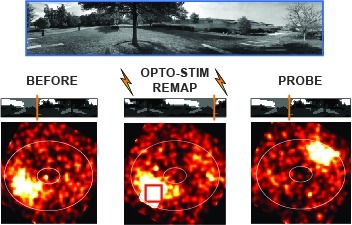
Many animals rely on an internal heading representation when navigating in varied environments. How this representation is linked to the sensory cues that define different surroundings is unclear. In the fly brain, heading is represented by 'compass' neurons that innervate a ring-shaped structure known as the ellipsoid body. Each compass neuron receives inputs from 'ring' neurons that are selective for particular visual features; this combination provides an ideal substrate for the extraction of directional information from a visual scene. Here we combine two-photon calcium imaging and optogenetics in tethered flying flies with circuit modelling, and show how the correlated activity of compass and visual neurons drives plasticity, which flexibly transforms two-dimensional visual cues into a stable heading representation. We also describe how this plasticity enables the fly to convert a partial heading representation, established from orienting within part of a novel setting, into a complete heading representation. Our results provide mechanistic insight into the memory-related computations that are essential for flexible navigation in varied surroundings.