Filter
Associated Lab
- Ahrens Lab (5) Apply Ahrens Lab filter
- Beyene Lab (1) Apply Beyene Lab filter
- Branson Lab (3) Apply Branson Lab filter
- Card Lab (1) Apply Card Lab filter
- Cardona Lab (1) Apply Cardona Lab filter
- Druckmann Lab (1) Apply Druckmann Lab filter
- Freeman Lab (4) Apply Freeman Lab filter
- Funke Lab (2) Apply Funke Lab filter
- Harris Lab (1) Apply Harris Lab filter
- Ji Lab (1) Apply Ji Lab filter
- Remove Keller Lab filter Keller Lab
- Lavis Lab (3) Apply Lavis Lab filter
- Liu (Zhe) Lab (1) Apply Liu (Zhe) Lab filter
- Looger Lab (5) Apply Looger Lab filter
- Pavlopoulos Lab (1) Apply Pavlopoulos Lab filter
- Schreiter Lab (1) Apply Schreiter Lab filter
- Stringer Lab (1) Apply Stringer Lab filter
- Tillberg Lab (1) Apply Tillberg Lab filter
- Turaga Lab (1) Apply Turaga Lab filter
- Turner Lab (1) Apply Turner Lab filter
Associated Project Team
Publication Date
- 2024 (2) Apply 2024 filter
- 2023 (1) Apply 2023 filter
- 2022 (1) Apply 2022 filter
- 2021 (2) Apply 2021 filter
- 2020 (5) Apply 2020 filter
- 2019 (6) Apply 2019 filter
- 2018 (6) Apply 2018 filter
- 2017 (2) Apply 2017 filter
- 2016 (6) Apply 2016 filter
- 2015 (8) Apply 2015 filter
- 2014 (8) Apply 2014 filter
- 2013 (10) Apply 2013 filter
- 2012 (3) Apply 2012 filter
- 2011 (4) Apply 2011 filter
- 2010 (4) Apply 2010 filter
- 2009 (1) Apply 2009 filter
- 2008 (3) Apply 2008 filter
- 2007 (1) Apply 2007 filter
- 2006 (2) Apply 2006 filter
- 2005 (1) Apply 2005 filter
Type of Publication
76 Publications
Showing 1-10 of 76 resultsGenetically encoded fluorescent calcium indicators allow cellular-resolution recording of physiology. However, bright, genetically targetable indicators that can be multiplexed with existing tools in vivo are needed for simultaneous imaging of multiple signals. Here we describe WHaloCaMP, a modular chemigenetic calcium indicator built from bright dye-ligands and protein sensor domains. Fluorescence change in WHaloCaMP results from reversible quenching of the bound dye via a strategically placed tryptophan. WHaloCaMP is compatible with rhodamine dye-ligands that fluoresce from green to near-infrared, including several that efficiently label the brain in animals. When bound to a near-infrared dye-ligand, WHaloCaMP shows a 7× increase in fluorescence intensity and a 2.1-ns increase in fluorescence lifetime upon calcium binding. We use WHaloCaMP1a to image Ca responses in vivo in flies and mice, to perform three-color multiplexed functional imaging of hundreds of neurons and astrocytes in zebrafish larvae and to quantify Ca concentration using fluorescence lifetime imaging microscopy (FLIM).
Understanding the diversification of mammalian cell lineages is an essential to embryonic development, organ regeneration and tissue engineering. Shortly after implantation in the uterus, the pluripotent cells of the mammalian epiblast generate the three germ layers: ectoderm, mesoderm and endoderm1. Although clonal analyses suggest early specification of epiblast cells towards particular cell lineages2–4, single-cell transcriptomes do not identify lineage-specific markers in the epiblast5–11 and thus, the molecular regulation of such specification remains unknow. Here, we studied the epigenetic landscape of single epiblast cells, which revealed lineage priming towards endoderm, ectoderm or mesoderm. Unexpectedly, epiblast cells with mesodermal priming show a strong signature for the endothelial/endocardial fate, suggesting early specification of this lineage aside from other mesoderm. Through clonal analysis and live imaging, we show that endothelial precursors show early lineage divergence from the rest of mesodermal derivatives. In particular, cardiomyocytes and endocardial cells show limited lineage relationship, despite being temporally and spatially co-recruited during gastrulation. Furthermore, analysing the live tracks of single cells through unsupervised classification of cell migratory activity, we found early behavioral divergence of endothelial precursors shortly after the onset of mesoderm migration towards the cardiogenic area. These results provide a new model for the phenotypically silent specification of mammalian cell lineages in pluripotent cells of the epiblast and modify current knowledge on the sequence and timing of cardiovascular lineages diversification.
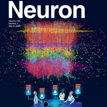
We present a method to automatically identify and track nuclei in time-lapse microscopy recordings of entire developing embryos. The method combines deep learning and global optimization. On a mouse dataset, it reconstructs 75.8% of cell lineages spanning 1 h, as compared to 31.8% for the competing method. Our approach improves understanding of where and when cell fate decisions are made in developing embryos, tissues, and organs.

We present a method to automatically identify and track nuclei in time-lapse microscopy recordings of entire developing embryos. The method combines deep learning and global optimization. On a mouse dataset, it reconstructs 75.8% of cell lineages spanning 1 h, as compared to 31.8% for the competing method. Our approach improves understanding of where and when cell fate decisions are made in developing embryos, tissues, and organs.

Glucose is arguably the most important molecule in metabolism, and its dysregulation underlies diabetes. We describe a family of single-wavelength genetically encoded glucose sensors with a high signal-to-noise ratio, fast kinetics, and affinities varying over four orders of magnitude (1 μM to 10 mM). The sensors allow mechanistic characterization of glucose transporters expressed in cultured cells with high spatial and temporal resolution. Imaging of neuron/glia co-cultures revealed ∼3-fold faster glucose changes in astrocytes. In larval Drosophila central nervous system explants, intracellular neuronal glucose fluxes suggested a rostro-caudal transport pathway in the ventral nerve cord neuropil. In zebrafish, expected glucose-related physiological sequelae of insulin and epinephrine treatments were directly visualized. Additionally, spontaneous muscle twitches induced glucose uptake in muscle, and sensory and pharmacological perturbations produced large changes in the brain. These sensors will enable rapid, high-resolution imaging of glucose influx, efflux, and metabolism in behaving animals.
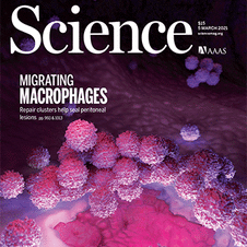
The mammalian heart is derived from multiple cell lineages; however, our understanding of when and how the diverse cardiac cell types arise is limited. We mapped the origin of the embryonic mouse heart at single-cell resolution using a combination of transcriptomic, imaging, and genetic lineage labeling approaches. This provided a transcriptional and anatomic definition of cardiac progenitor types. Furthermore, it revealed a cardiac progenitor pool that is anatomically and transcriptionally distinct from currently known cardiac progenitors. Besides contributing to cardiomyocytes, these cells also represent the earliest progenitor of the epicardium, a source of trophic factors and cells during cardiac development and injury. This study provides detailed insights into the formation of early cardiac cell types, with particular relevance to the development of cell-based cardiac regenerative therapies.

An important question in early neural development is the origin of stochastic nuclear movement between apical and basal surfaces of neuroepithelia during interkinetic nuclear migration. Tracking of nuclear subpopulations has shown evidence of diffusion - mean squared displacements growing linearly in time - and suggested crowding from cell division at the apical surface drives basalward motion. Yet, this hypothesis has not yet been tested, and the forces involved not quantified. We employ long-term, rapid light-sheet and two-photon imaging of early zebrafish retinogenesis to track entire populations of nuclei within the tissue. The time-varying concentration profiles show clear evidence of crowding as nuclei reach close-packing and are quantitatively described by a nonlinear diffusion model. Considerations of nuclear motion constrained inside the enveloping cell membrane show that concentration-dependent stochastic forces inside cells, compatible in magnitude to those found in cytoskeletal transport, can explain the observed magnitude of the diffusion constant.
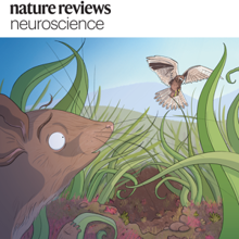
Tissue clearing and light-sheet microscopy have a 100-year-plus history, yet these fields have been combined only recently to facilitate novel experiments and measurements in neuroscience. Since tissue-clearing methods were first combined with modernized light-sheet microscopy a decade ago, the performance of both technologies has rapidly improved, broadening their applications. Here, we review the state of the art of tissue-clearing methods and light-sheet microscopy and discuss applications of these techniques in profiling cells and circuits in mice. We examine outstanding challenges and future opportunities for expanding these techniques to achieve brain-wide profiling of cells and circuits in primates and humans. Such integration will help provide a systems-level understanding of the physiology and pathology of our central nervous system.
Tissue clearing and light-sheet microscopy have a 100-year-plus history, yet these fields have been combined only recently to facilitate novel experiments and measurements in neuroscience. Since tissue-clearing methods were first combined with modernized light-sheet microscopy a decade ago, the performance of both technologies has rapidly improved, broadening their applications. Here, we review the state of the art of tissue-clearing methods and light-sheet microscopy and discuss applications of these techniques in profiling cells and circuits in mice. We examine outstanding challenges and future opportunities for expanding these techniques to achieve brain-wide profiling of cells and circuits in primates and humans. Such integration will help provide a systems-level understanding of the physiology and pathology of our central nervous system.
State-of-the-art tissue-clearing methods provide subcellular-level optical access to intact tissues from individual organs and even to some entire mammals. When combined with light-sheet microscopy and automated approaches to image analysis, existing tissue-clearing methods can speed up and may reduce the cost of conventional histology by several orders of magnitude. In addition, tissue-clearing chemistry allows whole-organ antibody labelling, which can be applied even to thick human tissues. By combining the most powerful labelling, clearing, imaging and data-analysis tools, scientists are extracting structural and functional cellular and subcellular information on complex mammalian bodies and large human specimens at an accelerated pace. The rapid generation of terabyte-scale imaging data furthermore creates a high demand for efficient computational approaches that tackle challenges in large-scale data analysis and management. In this Review, we discuss how tissue-clearing methods could provide an unbiased, system-level view of mammalian bodies and human specimens and discuss future opportunities for the use of these methods in human neuroscience.