Filter
Associated Lab
- Ahrens Lab (3) Apply Ahrens Lab filter
- Betzig Lab (8) Apply Betzig Lab filter
- Beyene Lab (1) Apply Beyene Lab filter
- Clapham Lab (3) Apply Clapham Lab filter
- Dudman Lab (2) Apply Dudman Lab filter
- Harris Lab (3) Apply Harris Lab filter
- Hess Lab (2) Apply Hess Lab filter
- Ji Lab (1) Apply Ji Lab filter
- Keller Lab (3) Apply Keller Lab filter
- Remove Lavis Lab filter Lavis Lab
- Lippincott-Schwartz Lab (7) Apply Lippincott-Schwartz Lab filter
- Liu (Zhe) Lab (15) Apply Liu (Zhe) Lab filter
- Looger Lab (8) Apply Looger Lab filter
- Pedram Lab (1) Apply Pedram Lab filter
- Podgorski Lab (2) Apply Podgorski Lab filter
- Schreiter Lab (8) Apply Schreiter Lab filter
- Shroff Lab (1) Apply Shroff Lab filter
- Singer Lab (6) Apply Singer Lab filter
- Spruston Lab (2) Apply Spruston Lab filter
- Stern Lab (2) Apply Stern Lab filter
- Sternson Lab (1) Apply Sternson Lab filter
- Stringer Lab (1) Apply Stringer Lab filter
- Svoboda Lab (4) Apply Svoboda Lab filter
- Tebo Lab (1) Apply Tebo Lab filter
- Tillberg Lab (2) Apply Tillberg Lab filter
- Tjian Lab (5) Apply Tjian Lab filter
- Turner Lab (3) Apply Turner Lab filter
- Wang (Shaohe) Lab (1) Apply Wang (Shaohe) Lab filter
Associated Project Team
Publication Date
- 2025 (11) Apply 2025 filter
- 2024 (13) Apply 2024 filter
- 2023 (9) Apply 2023 filter
- 2022 (13) Apply 2022 filter
- 2021 (10) Apply 2021 filter
- 2020 (9) Apply 2020 filter
- 2019 (6) Apply 2019 filter
- 2018 (12) Apply 2018 filter
- 2017 (16) Apply 2017 filter
- 2016 (13) Apply 2016 filter
- 2015 (5) Apply 2015 filter
- 2014 (7) Apply 2014 filter
- 2013 (4) Apply 2013 filter
- 2012 (4) Apply 2012 filter
- 2011 (5) Apply 2011 filter
- 2010 (1) Apply 2010 filter
- 2009 (2) Apply 2009 filter
- 2008 (4) Apply 2008 filter
- 2007 (3) Apply 2007 filter
- 2006 (2) Apply 2006 filter
Type of Publication
149 Publications
Showing 111-120 of 149 resultsFluorogenic molecules are important tools for biological and biochemical research. The majority of fluorogenic compounds have a simple input-output relationship, where a single chemical input yields a fluorescent output. Development of new systems where multiple inputs converge to yield an optical signal could refine and extend fluorogenic compounds by allowing greater spatiotemporal control over the fluorescent signal. Here, we introduce a new red-shifted fluorescein derivative, Virginia Orange, as an exceptional scaffold for single- and dual-input fluorogenic molecules. Unlike fluorescein, installation of a single masking group on Virginia Orange is sufficient to fully suppress fluorescence, allowing preparation of fluorogenic enzyme substrates with rapid, single-hit kinetics. Virginia Orange can also be masked with two independent moieties; both of these masking groups must be removed to induce fluorescence. This allows facile construction of multi-input fluorogenic probes for sophisticated sensing regimes and genetic targeting of latent fluorophores to specific cellular populations.
The structure of axonal arbors controls how signals from individual neurons are routed within the mammalian brain. However, the arbors of very few long-range projection neurons have been reconstructed in their entirety, as axons with diameters as small as 100 nm arborize in target regions dispersed over many millimeters of tissue. We introduce a platform for high-resolution, three-dimensional fluorescence imaging of complete tissue volumes that enables the visualization and reconstruction of long-range axonal arbors. This platform relies on a high-speed two-photon microscope integrated with a tissue vibratome and a suite of computational tools for large-scale image data. We demonstrate the power of this approach by reconstructing the axonal arbors of multiple neurons in the motor cortex across a single mouse brain.
The rhodamine system is a flexible framework for building small-molecule fluorescent probes. Changing N-substitution patterns and replacing the xanthene oxygen with a dimethylsilicon moiety can shift the absorption and fluorescence emission maxima of rhodamine dyes to longer wavelengths. Acylation of the rhodamine nitrogen atoms forces the molecule to adopt a nonfluorescent lactone form, providing a convenient method to make fluorogenic compounds. Herein, we take advantage of all of these structural manipulations and describe a novel photoactivatable fluorophore based on a Si-containing analogue of Q-rhodamine. This probe is the first example of a "caged" Si-rhodamine, exhibits higher photon counts compared to established localization microscopy dyes, and is sufficiently red-shifted to allow multicolor imaging. The dye is a useful label for super-resolution imaging and constitutes a new scaffold for far-red fluorogenic molecules.
Observation of molecular processes inside living cells is fundamental to a quantitative understanding of how biological systems function. Specifically, decoding the complex behavior of single molecules enables us to measure kinetics, transport, and self-assembly at this fundamental level that is often veiled in ensemble experiments. In the past decade, rapid developments in fluorescence microscopy, fluorescence correlation spectroscopy, and fluorescent labeling techniques have enabled new experiments to investigate the robustness and stochasticity of diverse molecular mechanisms with high spatiotemporal resolution. This review discusses the concepts and strategies of structural and functional imaging in living cells at the single-molecule level with minimal perturbations to the specimen.
Enzyme kinetics measurements are a standard component of undergraduate biochemistry laboratories. The combination of serine hydrolases and fluorogenic enzyme substrates provides a rapid, sensitive, and general method for measuring enzyme kinetics in an undergraduate biochemistry laboratory. In this method, the kinetic activity of multiple protein variants is determined in parallel using a microplate reader, multichannel pipets, serial dilutions, and fluorogenic ester substrates. The utility of this methodology is illustrated by the measurement of differential enzyme activity in microplate volumes in triplicate with small protein samples and low activity enzyme variants. Enzyme kinetic measurements using fluorogenic substrates are, thus, adaptable for use with student-purified enzyme variants and for comparative enzyme kinetics studies. The rapid setup and analysis of these kinetic experiments not only provides advanced undergraduates with experience in a fundamental biochemical technique, but also provides the adaptability for use in inquiry-based laboratories.
Specific labeling of biomolecules with bright fluorophores is the keystone of fluorescence microscopy. Genetically encoded self-labeling tag proteins can be coupled to synthetic dyes inside living cells, resulting in brighter reporters than fluorescent proteins. Intracellular labeling using these techniques requires cell-permeable fluorescent ligands, however, limiting utility to a small number of classic fluorophores. Here we describe a simple structural modification that improves the brightness and photostability of dyes while preserving spectral properties and cell permeability. Inspired by molecular modeling, we replaced the N,N-dimethylamino substituents in tetramethylrhodamine with four-membered azetidine rings. This addition of two carbon atoms doubles the quantum efficiency and improves the photon yield of the dye in applications ranging from in vitro single-molecule measurements to super-resolution imaging. The novel substitution is generalizable, yielding a palette of chemical dyes with improved quantum efficiencies that spans the UV and visible range.
Combinatorial cis-regulatory networks encoded in animal genomes represent the foundational gene expression mechanism for directing cell-fate commitment and maintenance of cell identity by transcription factors (TFs). However, the 3D spatial organization of cis-elements and how such sub-nuclear structures influence TF activity remain poorly understood. Here, we combine lattice light-sheet imaging, single-molecule tracking, numerical simulations, and ChIP-exo mapping to localize and functionally probe Sox2 enhancer-organization in living embryonic stem cells. Sox2 enhancers form 3D-clusters that are segregated from heterochromatin but overlap with a subset of Pol II enriched regions. Sox2 searches for specific binding targets via a 3D-diffusion dominant mode when shuttling long-distances between clusters while chromatin-bound states predominate within individual clusters. Thus, enhancer clustering may reduce global search efficiency but enables rapid local fine-tuning of TF search parameters. Our results suggest an integrated model linking cis-element 3D spatial distribution to local-versus-global target search modalities essential for regulating eukaryotic gene transcription.
Pheromones, chemical signals that convey social information, mediate many insect social behaviors, including navigation and aggregation. Several studies have suggested that behavior during the immature larval stages of Drosophila development is influenced by pheromones, but none of these compounds or the pheromone-receptor neurons that sense them have been identified. Here we report a larval pheromone-signaling pathway. We found that larvae produce two novel long-chain fatty acids that are attractive to other larvae. We identified a single larval chemosensory neuron that detects these molecules. Two members of the pickpocket family of DEG/ENaC channel subunits (ppk23 and ppk29) are required to respond to these pheromones. This pheromone system is evolving quickly, since the larval exudates of D. simulans, the sister species of D. melanogaster, are not attractive to other larvae. Our results define a new pheromone signaling system in Drosophila that shares characteristics with pheromone systems in a wide diversity of insects.
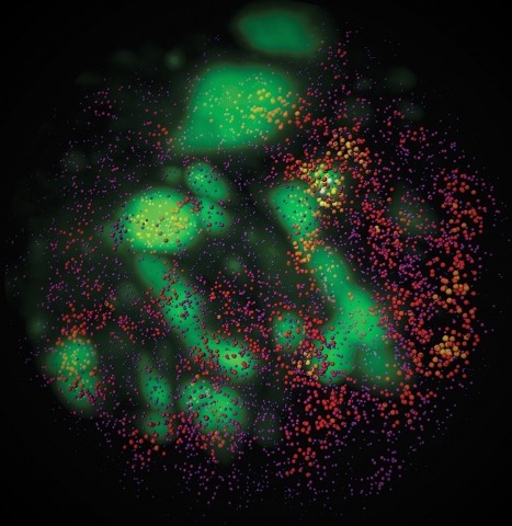
The molecular and cellular architecture of the organs in a whole mouse is revealed through optical clearing.


