Filter
Associated Lab
- Ahrens Lab (2) Apply Ahrens Lab filter
- Betzig Lab (9) Apply Betzig Lab filter
- Hess Lab (1) Apply Hess Lab filter
- Ji Lab (2) Apply Ji Lab filter
- Lavis Lab (1) Apply Lavis Lab filter
- Pedram Lab (2) Apply Pedram Lab filter
- Saalfeld Lab (1) Apply Saalfeld Lab filter
- Remove Shroff Lab filter Shroff Lab
- Wang (Shaohe) Lab (2) Apply Wang (Shaohe) Lab filter
Associated Project Team
Publication Date
Type of Publication
31 Publications
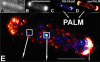
Showing 21-30 of 31 resultsRecent advances in optical microscopy have enabled biological imaging beyond the diffraction limit at nanometer resolution. A general feature of most of the techniques based on photoactivated localization microscopy (PALM) or stochastic optical reconstruction microscopy (STORM) has been the use of thin biological samples in combination with total internal reflection, thus limiting the imaging depth to a fraction of an optical wavelength. However, to study whole cells or organelles that are typically up to 15 microm deep into the cell, the extension of these methods to a three-dimensional (3D) super resolution technique is required. Here, we report an advance in optical microscopy that enables imaging of protein distributions in cells with a lateral localization precision better than 50 nm at multiple imaging planes deep in biological samples. The approach is based on combining the lateral super resolution provided by PALM with two-photon temporal focusing that provides optical sectioning. We have generated super-resolution images over an axial range of approximately 10 microm in both mitochondrially labeled fixed cells, and in the membranes of living S2 Drosophila cells.
Fluorescence microscopy is an invaluable tool in biology, yet its performance is compromised when the wavefront of light is distorted due to optical imperfections or the refractile nature of the sample. Such optical aberrations can dramatically lower the information content of images by degrading image contrast, resolution, and signal. Adaptive optics (AO) methods can sense and subsequently cancel the aberrated wavefront, but are too complex, inefficient, slow, or expensive for routine adoption by most labs. Here we introduce a rapid, sensitive, and robust wavefront sensing scheme based on phase diversity, a method successfully deployed in astronomy but underused in microscopy. Our method enables accurate wavefront sensing to less than λ/35 root mean square (RMS) error with few measurements, and AO with no additional hardware besides a corrective element. After validating the method with simulations, we demonstrate calibration of a deformable mirror > 100-fold faster than comparable methods (corresponding to wavefront sensing on the ~100 ms scale), and sensing and subsequent correction of severe aberrations (RMS wavefront distortion exceeding λ/2), restoring diffraction-limited imaging on extended biological samples.
Key to understanding a protein’s biological function is the accurate determination of its spatial distribution inside a cell. Although fluorescent protein markers allow the targeting of specific proteins with molecular precision, much of this information is lost when the resultant fusion proteins are imaged with conventional, diffraction-limited optics. In response, several imaging modalities that are capable of resolution below the diffraction limit (approximately 200 nm) have emerged. Here, both single- and dual-color superresolution imaging of biological structures using photoactivated localization microscopy (PALM) are described. The examples discussed focus on adhesion complexes: dense, protein-filled assemblies that form at the interface between cells and their substrata. A particular emphasis is placed on the instrumentation and photoactivatable fluorescent protein (PA-FP) tags necessary to achieve PALM images at approximately 20 nm resolution in 5 to 30 min in fixed cells.
Commentary: A paper spearheaded by Hari which gives a thorough description of the methods and hardware needed to successfully practice PALM, including cover slip preparation, cell transfection and fixation, drift correction with fiducials, characterization of on/off contrast ratios for different photoactivted fluorescent proteins, identifying PALM-suitable cells, and mechanical and optical components of a PALM system.
Determining cell identities in imaging sequences is an important yet challenging task. The conventional method for cell identification is via cell tracking, which is complex and can be time-consuming. In this study, we propose an innovative approach to cell identification during early C. elegans embryogenesis using machine learning. We employed random forest, MLP, and LSTM models, and tested cell classification accuracy on 3D time-lapse confocal datasets spanning the first 4 hours of embryogenesis. By leveraging a small number of spatial-temporal features of individual cells, including cell trajectory and cell fate information, our models achieve an accuracy of over 90%, even with limited data. We also determine the most important feature contributions and can interpret these features in the context of biological knowledge. Our research demonstrates the success of predicting cell identities in 4D imaging sequences directly from simple spatio-temporal features.
Super-resolution microscopy (SRM) is gaining popularity in biosciences; however, claims about optical resolution are contested and often misleading. In this Viewpoint, experts share their views on resolution and common trade-offs, such as labelling and post-processing, aiming to clarify them for biologists and facilitate deeper understanding and best use of SRM.
Super-resolution microscopy (SRM) has undeniable potential for scientific discovery, yet still presents many challenges that hinder its widespread adoption, including technical trade-offs between resolution, speed and photodamage, as well as limitations in imaging live samples and larger, more complex biological structures. Furthermore, SRM often requires specialized expertise and complex instrumentation, which can deter biologists from fully embracing the technology. In this Perspective, a follow-up to our recent Q&A article, we aim to demystify these challenges by addressing common questions and misconceptions surrounding SRM. Experts offer practical insights into how biologists can maximize the benefits of SRM while navigating issues such as photobleaching, image artifacts and the limitations of existing techniques. We also highlight recent developments in SRM that continue to push the boundaries of resolution. Our goal is to equip researchers with the crucial knowledge they need to harness the full potential of SRM.
The Escherichia coli chemotaxis network is a model system for biological signal processing. In E. coli, transmembrane receptors responsible for signal transduction assemble into large clusters containing several thousand proteins. These sensory clusters have been observed at cell poles and future division sites. Despite extensive study, it remains unclear how chemotaxis clusters form, what controls cluster size and density, and how the cellular location of clusters is robustly maintained in growing and dividing cells. Here, we use photoactivated localization microscopy (PALM) to map the cellular locations of three proteins central to bacterial chemotaxis (the Tar receptor, CheY, and CheW) with a precision of 15 nm. We find that cluster sizes are approximately exponentially distributed, with no characteristic cluster size. One-third of Tar receptors are part of smaller lateral clusters and not of the large polar clusters. Analysis of the relative cellular locations of 1.1 million individual proteins (from 326 cells) suggests that clusters form via stochastic self-assembly. The super-resolution PALM maps of E. coli receptors support the notion that stochastic self-assembly can create and maintain approximately periodic structures in biological membranes, without direct cytoskeletal involvement or active transport.
Commentary: Our goal as tool developers is to invent methods capable of uncovering new biological insights unobtainable by pre-existing technologies. A terrific example is given by this paper, where grad students Derek Greenfield and Ann McEvoy in Jan Liphardt’s group at Berkeley used our PALM to image the size and position distributions of chemotaxis proteins in E. Coli with unprecedented precision and sensitivity. Their analysis revealed that the cluster sizes follow a stretched exponential distribution, and the density of clusters is highest furthest away from the largest (e.g., polar) clusters. Both observations support a model for passive self-assembly rather than active cytoskeletal assembly of the chemotaxis network.
Within dendritic spines, actin is presumed to anchor receptors in the postsynaptic density and play numerous roles regulating synaptic transmission. However, the submicron dimensions of spines have hindered examination of actin dynamics within them and prevented live-cell discrimination of perisynaptic actin filaments. Using photoactivated localization microscopy, we measured movement of individual actin molecules within living spines. Velocity of single actin molecules along filaments, an index of filament polymerization rate, was highly heterogeneous within individual spines. Most strikingly, molecular velocity was elevated in discrete, well-separated foci occurring not principally at the spine tip, but in subdomains throughout the spine, including the neck. Whereas actin velocity on filaments at the synapse was substantially elevated, at the endocytic zone there was no enhanced polymerization activity. We conclude that actin subserves spatially diverse, independently regulated processes throughout spines. Perisynaptic actin forms a uniquely dynamic structure well suited for direct, active regulation of the synapse.
Commentary: A nice application of single particle tracking PALM (sptPALM), showing the flow of actin in the spines of live cultured neurons. Since 2008, the PALM in our lab has largely become a user facility, available to outside users as well as Janelians. Grad student Nick Frost in Tom Blanpied’s group at the U. of Maryland Med School visited on a number of occasions to use the PALM, with training and assistance from Hari.
We have demonstrated super-resolution imaging of protein distributions in cells at depth at multiple layers with a lateral localization precision better than 50 nm. The approach is based on combining photoactivated localization microscopy with temporal focusing.
Polarized fluorescence microscopy is a valuable tool for measuring molecular orientations, but techniques for recovering three-dimensional orientations and positions of fluorescent ensembles are limited. We report a polarized dual-view light-sheet system for determining the three-dimensional orientations and diffraction-limited positions of ensembles of fluorescent dipoles that label biological structures, and we share a set of visualization, histogram, and profiling tools for interpreting these positions and orientations. We model our samples, their excitation, and their detection using coarse-grained representations we call orientation distribution functions (ODFs). We apply ODFs to create physics-informed models of image formation with spatio-angular point-spread and transfer functions. We use theory and experiment to conclude that light-sheet tilting is a necessary part of our design for recovering all three-dimensional orientations. We use our system to extend known two-dimensional results to three dimensions in FM1-43-labelled giant unilamellar vesicles, fast-scarlet-labelled cellulose in xylem cells, and phalloidin-labelled actin in U2OS cells. Additionally, we observe phalloidin-labelled actin in mouse fibroblasts grown on grids of labelled nanowires and identify correlations between local actin alignment and global cell-scale orientation, indicating cellular coordination across length scales.Competing Interest StatementH.S., A.K., S.M., P.L.R., R.O., Y.W., and T.C. hold US Patent #11428632.