Filter
Associated Lab
- Aso Lab (1) Apply Aso Lab filter
- Remove Betzig Lab filter Betzig Lab
- Bock Lab (1) Apply Bock Lab filter
- Clapham Lab (2) Apply Clapham Lab filter
- Fetter Lab (2) Apply Fetter Lab filter
- Harris Lab (2) Apply Harris Lab filter
- Hess Lab (6) Apply Hess Lab filter
- Ji Lab (11) Apply Ji Lab filter
- Lavis Lab (8) Apply Lavis Lab filter
- Lippincott-Schwartz Lab (6) Apply Lippincott-Schwartz Lab filter
- Liu (Zhe) Lab (7) Apply Liu (Zhe) Lab filter
- Magee Lab (2) Apply Magee Lab filter
- Rubin Lab (1) Apply Rubin Lab filter
- Saalfeld Lab (2) Apply Saalfeld Lab filter
- Schreiter Lab (1) Apply Schreiter Lab filter
- Shroff Lab (9) Apply Shroff Lab filter
- Singer Lab (1) Apply Singer Lab filter
- Svoboda Lab (2) Apply Svoboda Lab filter
- Tjian Lab (4) Apply Tjian Lab filter
- Turner Lab (1) Apply Turner Lab filter
Associated Project Team
Publication Date
- 2025 (2) Apply 2025 filter
- 2024 (2) Apply 2024 filter
- 2023 (4) Apply 2023 filter
- 2022 (3) Apply 2022 filter
- 2021 (2) Apply 2021 filter
- 2020 (4) Apply 2020 filter
- 2019 (7) Apply 2019 filter
- 2018 (6) Apply 2018 filter
- 2017 (8) Apply 2017 filter
- 2016 (12) Apply 2016 filter
- 2015 (11) Apply 2015 filter
- 2014 (8) Apply 2014 filter
- 2013 (4) Apply 2013 filter
- 2012 (5) Apply 2012 filter
- 2011 (7) Apply 2011 filter
- 2010 (3) Apply 2010 filter
- 2009 (2) Apply 2009 filter
- 2008 (8) Apply 2008 filter
- 2007 (2) Apply 2007 filter
- 2006 (1) Apply 2006 filter
Type of Publication
- Remove Janelia filter Janelia
101 Publications
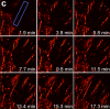
Showing 61-70 of 101 resultsThe inner ear is a fluid-filled closed-epithelial structure whose function requires maintenance of an internal hydrostatic pressure and fluid composition. The endolymphatic sac (ES) is a dead-end epithelial tube connected to the inner ear whose function is unclear. ES defects can cause distended ear tissue, a pathology often seen in hearing and balance disorders. Using live imaging of zebrafish larvae, we reveal that the ES undergoes cycles of slow pressure-driven inflation followed by rapid deflation. Absence of these cycles in mutants leads to distended ear tissue. Using serial-section electron microscopy and adaptive optics lattice light-sheet microscopy, we find a pressure relief valve in the ES comprised of partially separated apical junctions and dynamic overlapping basal lamellae that separate under pressure to release fluid. We propose that this lmx1-dependent pressure relief valve is required to maintain fluid homeostasis in the inner ear and other fluid-filled cavities.
Although fluorescence microscopy provides a crucial window into the physiology of living specimens, many biological processes are too fragile, are too small, or occur too rapidly to see clearly with existing tools. We crafted ultrathin light sheets from two-dimensional optical lattices that allowed us to image three-dimensional (3D) dynamics for hundreds of volumes, often at subsecond intervals, at the diffraction limit and beyond. We applied this to systems spanning four orders of magnitude in space and time, including the diffusion of single transcription factor molecules in stem cell spheroids, the dynamic instability of mitotic microtubules, the immunological synapse, neutrophil motility in a 3D matrix, and embryogenesis in Caenorhabditis elegans and Drosophila melanogaster. The results provide a visceral reminder of the beauty and the complexity of living systems.
We demonstrate live-cell super-resolution imaging using photoactivated localization microscopy (PALM). The use of photon-tolerant cell lines in combination with the high resolution and molecular sensitivity of PALM permitted us to investigate the nanoscale dynamics within individual adhesion complexes (ACs) in living cells under physiological conditions for as long as 25 min, with half of the time spent collecting the PALM images at spatial resolutions down to approximately 60 nm and frame rates as short as 25 s. We visualized the formation of ACs and measured the fractional gain and loss of individual paxillin molecules as each AC evolved. By allowing observation of a wide variety of nanoscale dynamics, live-cell PALM provides insights into molecular assembly during the initiation, maturation and dissolution of cellular processes.
Commentary: The first example of true live cell and time lapse imaging by localization microscopy (as opposed to particle tracking), this paper uses the Nyquist criterion to establish a necessary condition for true spatial resolution based on the density of localized molecules – a condition often unmet in claims elsewhere in the superresolution literature.
By any method, higher spatiotemporal resolution requires increasing light exposure at the specimen, making noninvasive imaging increasingly difficult. Here, simultaneous differential interference contrast imaging is used to establish that cells behave physiologically before, during, and after PALM imaging. Similar controls are lacking from many supposed “live cell” superresolution demonstrations.
Membrane remodeling is an essential part for transfer of components to and from the cell surface and membrane-bound organelles, and for changes in cell shape, particularly critical during cell division. Earlier analyses, based on classical optical live-cell imaging, mostly restricted by technical necessity to the attached bottom surface, showed persistent formation of endocytic clathrin pits and vesicles during mitosis. Taking advantage of the resolution, speed, and non-invasive illumination of the newly developed lattice light sheet fluorescence microscope, we reexamined their assembly dynamics over the entire cell surface and showed that clathrin pits form at a lower rate during late mitosis. Full-cell imaging measurements of cell surface area and volume throughout the cell cycle of single cells in culture and in zebrafish embryos showed that the total surface increased rapidly during the transition from telophase to cytokinesis, whereas cell volume increased slightly in metaphase and remained relatively constant during cytokinesis. These applications demonstrate the advantage of lattice light sheet microscopy and enable a new standard for imaging membrane dynamics in single cells and in multicellular assemblies.
Recent methods have revealed that cells on planar substrates exert both shear (in-plane) and normal (out-of-plane) tractions against the extracellular matrix (ECM). However, the location and origin of the normal tractions with respect to the adhesive and cytoskeletal elements of cells have not been elucidated. We developed a high-spatiotemporal-resolution, multidimensional (2.5D) traction force microscopy to measure and model the full 3D nature of cellular forces on planar 2D surfaces. We show that shear tractions are centered under elongated focal adhesions whereas upward and downward normal tractions are detected on distal (toward the cell edge) and proximal (toward the cell body) ends of adhesions, respectively. Together, these forces produce significant rotational moments about focal adhesions in both protruding and retracting peripheral regions. Temporal 2.5D traction force microscopy analysis of migrating and spreading cells shows that these rotational moments are highly dynamic, propagating outward with the leading edge of the cell. Finally, we developed a finite element model to examine how rotational moments could be generated about focal adhesions in a thin lamella. Our model suggests that rotational moments can be generated largely via shear lag transfer to the underlying ECM from actomyosin contractility applied at the intracellular surface of a rigid adhesion of finite thickness. Together, these data demonstrate and probe the origin of a previously unappreciated multidimensional stress profile associated with adhesions and highlight the importance of new approaches to characterize cellular forces.
Recent advances in optical microscopy have enabled biological imaging beyond the diffraction limit at nanometer resolution. A general feature of most of the techniques based on photoactivated localization microscopy (PALM) or stochastic optical reconstruction microscopy (STORM) has been the use of thin biological samples in combination with total internal reflection, thus limiting the imaging depth to a fraction of an optical wavelength. However, to study whole cells or organelles that are typically up to 15 microm deep into the cell, the extension of these methods to a three-dimensional (3D) super resolution technique is required. Here, we report an advance in optical microscopy that enables imaging of protein distributions in cells with a lateral localization precision better than 50 nm at multiple imaging planes deep in biological samples. The approach is based on combining the lateral super resolution provided by PALM with two-photon temporal focusing that provides optical sectioning. We have generated super-resolution images over an axial range of approximately 10 microm in both mitochondrially labeled fixed cells, and in the membranes of living S2 Drosophila cells.
Class-18 myosins are most closely related to conventional class-2 nonmuscle myosins (NM2). Surprisingly, the purified head domains of Drosophila, mouse, and human myosin 18A (M18A) lack actin-activated ATPase activity and the ability to translocate actin filaments, suggesting that the functions of M18A in vivo do not depend on intrinsic motor activity. M18A has the longest coiled coil of any myosin outside of the class-2 myosins, suggesting that it might form bipolar filaments similar to conventional myosins. To address this possibility, we expressed and purified full-length mouse M18A using the baculovirus/Sf9 system. M18A did not form large bipolar filaments under any of the conditions tested. Instead, M18A formed an ∼65-nm-long bipolar structure with two heads at each end. Importantly, when NM2 was polymerized in the presence of M18A, the two myosins formed mixed bipolar filaments, as evidenced by cosedimentation, electron microscopy, and single-molecule imaging. Moreover, super-resolution imaging of NM2 and M18A using fluorescently tagged proteins and immunostaining of endogenous proteins showed that NM2 and M18A are present together within individual filaments inside living cells. Together, our in vitro and live-cell imaging data argue strongly that M18A coassembles with NM2 into mixed bipolar filaments. M18A could regulate the biophysical properties of these filaments and, by virtue of its extra N- and C-terminal domains, determine the localization and/or molecular interactions of the filaments. Given the numerous, fundamental cellular and developmental roles attributed to NM2, our results have far-reaching biological implications.
Retinal Müller glia function as injury-induced stem-like cells in zebrafish but not mammals. However, insights gleaned from zebrafish have been applied to stimulate nascent regenerative responses in the mammalian retina. For instance, microglia/macrophages regulate Müller glia stem cell activity in the chick, zebrafish, and mouse. We previously showed that post-injury immunosuppression by the glucocorticoid dexamethasone accelerated retinal regeneration kinetics in zebrafish. Similarly, microglia ablation enhances regenerative outcomes in the mouse retina. Targeted immunomodulation of microglia reactivity may therefore enhance the regenerative potential of Müller glia for therapeutic purposes. Here, we investigated potential mechanisms by which post-injury dexamethasone accelerates retinal regeneration kinetics, and the effects of dendrimer-based targeting of dexamethasone to reactive microglia. Intravital time-lapse imaging revealed that post-injury dexamethasone inhibited microglia reactivity. The dendrimer-conjugated formulation: (1) decreased dexamethasone-associated systemic toxicity, (2) targeted dexamethasone to reactive microglia, and (3) improved the regeneration enhancing effects of immunosuppression by increasing stem/progenitor proliferation rates. Lastly, we show that the gene rnf2 is required for the enhanced regeneration effect of D-Dex. These data support the use of dendrimer-based targeting of reactive immune cells to reduce toxicity and enhance the regeneration promoting effects of immunosuppressants in the retina.
Optical imaging of the dynamics of living specimens involves tradeoffs between spatial resolution, temporal resolution, and phototoxicity, made more difficult in three dimensions. Here, however, we report that rapid three-dimensional (3D) dynamics can be studied beyond the diffraction limit in thick or densely fluorescent living specimens over many time points by combining ultrathin planar illumination produced by scanned Bessel beams with super-resolution structured illumination microscopy. We demonstrate in vivo karyotyping of chromosomes during mitosis and identify different dynamics for the actin cytoskeleton at the dorsal and ventral surfaces of fibroblasts. Compared to spinning disk confocal microscopy, we demonstrate substantially reduced photodamage when imaging rapid morphological changes in D. discoideum cells, as well as improved contrast and resolution at depth within developing C. elegans embryos. Bessel beam structured plane illumination thus promises new insights into complex biological phenomena that require 4D subcellular spatiotemporal detail in either a single or multicellular context.
Nonmuscle myosin II (NM II) powers myriad developmental and cellular processes, including embryogenesis, cell migration, and cytokinesis [1]. To exert its functions, monomers of NM II assemble into bipolar filaments that produce a contractile force on the actin cytoskeleton. Mammalian cells express up to three isoforms of NM II (NM IIA, IIB, and IIC), each of which possesses distinct biophysical properties and supports unique as well as redundant cellular functions [2-8]. Despite previous efforts [9-13], it remains unclear whether NM II isoforms assemble in living cells to produce mixed (heterotypic) bipolar filaments or whether filaments consist entirely of a single isoform (homotypic). We addressed this question using fluorescently tagged versions of NM IIA, IIB, and IIC, isoform-specific immunostaining of the endogenous proteins, and two-color total internal reflection fluorescence structured-illumination microscopy, or TIRF-SIM, to visualize individual myosin II bipolar filaments inside cells. We show that NM II isoforms coassemble into heterotypic filaments in a variety of settings, including various types of stress fibers, individual filaments throughout the cell, and the contractile ring. We also show that the differential distribution of NM IIA and NM IIB typically seen in confocal micrographs of well-polarized cells is reflected in the composition of individual bipolar filaments. Interestingly, this differential distribution is less pronounced in freshly spread cells, arguing for the existence of a sorting mechanism acting over time. Together, our work argues that individual NM II isoforms are potentially performing both isoform-specific and isoform-redundant functions while coassembled with other NM II isoforms.