In addition to providing consultation and support, including help with writing M&M sections and Acknowledgements, other resources at the facility include multiple computer workstations with installed software for image visualization and analysis, a sample preparation laboratory, and supplementary microscope objectives.
Image Analysis Support
The facility offers free consultation on experiment design as well as image visualization and processing; please contact us in advance as suitable acquisition settings are a prerequisite for accurate analysis and quantification. We can also provide comprehensive image and data analysis support for multiple software packages through hands-on assistance and/or custom-written macros/plugins/scripts for ImageJ/FIJI, MATLAB, Imaris, etc.
Image Analysis: What method is right for you and how can we help?
What is the underlying biological hypothesis? Interaction? Co-expression? Proximity? Volume Overlap? The facility has several tools and experiences to quantify and visualize biological phenomenon. We can support the following image analysis modalities found below. If you have interests in additional readouts not listed, the Light Microscopy Facility can work with you to best find a method.

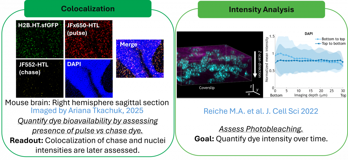

Image Analysis:Tools we Have
To accomplish the above analysis modalities, our workstations are installed with Amira, CellProfiler, ImageJ, Imaris, MATLAB, and Strataquest.
For a full list of additional software on our workstations please refer to our Image Analysis Software and Programming Tools section.
Image Analysis: Statistical Services
The Light Microscopy Facility can provide consultation on statistical and functional imaging analysis.
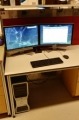
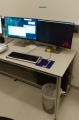
Computer Workstations
We have several workstations dedicated to viewing and processing of large, image datasets acquired with the facility's instruments, particularly the light sheet microscopes. A suite of imaging software (below), including a full version of Imaris, is installed on most of the workstations. Please contact us for assistance or to install additional software.
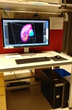
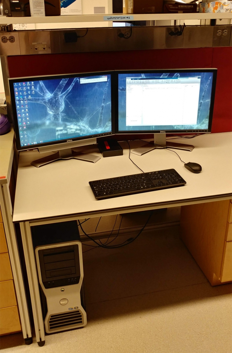
Workstation #1 (Imaris)

- Hardware:
- Dell Precision 7920, Windows 10 Enterprise 64-bit
- Intel Xeon Gold 6136 @3.0GHz, 512GB RAM (24 cores)
- Nvidia Quadro P6000 24GB GPU
- Storage: 8TB (SSD) and 10TB (HDD)
- Software Highlights:
Workstation #2
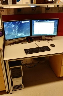
Workstation #3 (Strataquest)

- Hardware:
- Dell Precision 7920, Windows 10 Enterprise 64-bit
- Intel Xeon Gold 6242 @2.8GHz, 512GB RAM (32 cores)
- Nvidia Quadro RTX 5000 16GB GPU
- Storage: 2TB (SSD) and 6TB (HDD)
- Software Highlights:
- Strataquest, Imaris (Core), Imaris Stitcher, Amira, Vision4D (demo), Adobe CS5, Zen Lite, MATLAB, ImageJ/FIJI, MS Office, etc.
Workstation #4 (HIVE)
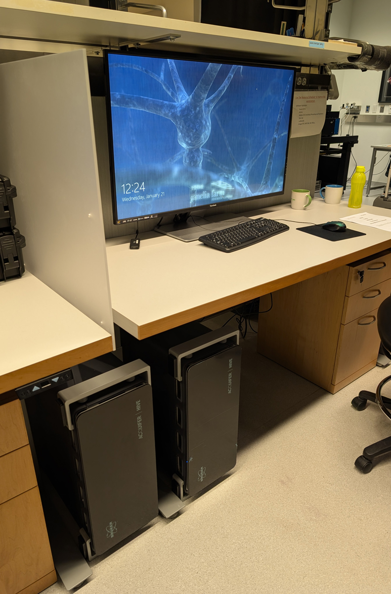
- Hardware:
- Acquifer HIVE L, Windows Server 2022 Standard 64-bit
- Intel Xeon Silver 4310 @2.10GHz, 1024GB RAM (24 cores)
- Nvidia RTX 5000 Ada 32GB GPU
- Storage: 12TB (SSD) and 150TB (HDD)
- Software Highlights:
Image Acquisition and Visualization Software
- Zen Blue and Zen Black: Software for controlling Zeiss widefield and laser scanning microscopes, respectively; they also serve as user-friendly image processing programs with methods for airyscan processing, spectral unmixing, batch processing, orthogonal projection, and stitching.
- Zen Lite: A free, stand-alone version for viewing .czi files with limited processing features.
- NIS-Elements AR: Software for controlling Nikon microscopes; it also serves as a user-friendly image processing program with tools for good 3D/4D visualization and High Content Analysis (HCA).
- NIS-Elements Viewer: A free, stand-alone version to view .nd2 files with limited processing features.
- cellSens: Image acquisition software designed for advanced image capture and control of motorized and encoded components on Olympus microscopes.
- IrfanView: A popular, free graphic viewer commonly used for digital microscopy slides.
- TissueFAXS Viewer: A stand-alone program for visualization of IHC/IF staining of digital slide images obtained with the TissueGnostics TissueFAXS 200 slide scanner.
- VLC: A free, cross-platform multimedia player that plays most multimedia files as well as DVDs, Audio CDs, and various streaming protocols.
- ChimeraX: A free, open-source molecular visualization program for graphics design.
- Agave: A free, open-source visualization tool for exploring and examining multichannel 3D volume data, including time series.
Image and Photo Manipulation Software
- Adobe Creative Suite 5 (CS5) Design Standard (includes Photoshop and Illustrator): Raster- and vector-based graphics editors, respectively, offering a variety of powerful tools with multiple image-editing functions. They are the de facto industry standard in photo manipulation and often used for publishing.
- GNU Image Manipulation Program (GIMP): A free, open-source, cross-platform image editor providing sophisticated tools along with 3rd party plugins for graphics design.
- Blender: A free, open-source 3D graphics software tool set for interactive applications.
Image Analysis Software and Programming Tools
- Imaris: Image visualization and analysis software capable of interactive 3D/4D rendering for large, multi-channel, microscopy datasets. Analysis includes cell counting, cell tracking, neuron tracing image segmentation, co-localization, and 4D movie generation. It has plugin capabilities for integrating custom-written macros from MATLAB, ImageJ, Python, etc.
- Imaris Viewer: A free, stand-alone version to view 3D datasets with limited processing features.
- Imaris Stitcher: A stand-alone application made for precise alignment and three-dimensional fusing of multiple microscopy image tiles.
- Amira: Image visualization and analysis software capable of interactive 3D/4D rendering for large, multi-channel, microscopy datasets. Analysis includes automatic and manual tools for enhanced 3D image segmentation and reconstruction using an interactive programming architecture.
- Arivis Pro: Image visualization and analysis software capable of interactive 3D/4D rendering for large microscopy datasets.
- Aivia: Image visualization with artificial intelligence-guided image analysis for data-driven scientific discovery.
- Napari: Image visualization and analysis software capable of interactive 3D/4D rendering for large microscopy datasets based in Python.
- Strataquest: Image visualization and analysis software for automated detection/cytometry and context-based quantification tasks across entire digital microscopy slides.
- Volocity: Image visualization and analysis software capable of colocalization and tracking for 3D/4D datasets.
- ImageJ: A popular free, open-source Java application that offers versatility for image processing via custom acquisition, analysis, and processing plugins.
- Icy: An open-community platform for bioimage informatics, providing software resources to visualize, annotate, and quantify bioimaging data with native ImageJ integration.
- CellProfiler: An open-source Python application developed to facilitate high content image analysis; no programming knowledge required.
- ilastik: An open-source Python application for image classification, segmentation, and analysis; no previous experience in image processing required.
- FluoRender: An open-source interactive rendering tool for attractive visual display of 3D/4D image datasets that also supports advanced segmentation.
- VVDviewer: An open-source application, built upon FluoRender, for visualization and analysis of 3D/4D datasets. It also supports advanced segmentation.
- Vaa3D: An open-source application for real-time 3D visualization and quantitative analysis of large-scale image datasets.
- MATLAB: A multi-paradigm, numerical computing environment and proprietary programming language that allows matrix manipulation, advanced plotting, implementation of algorithms, creation of user interfaces, etc. Multiple, specialized toolboxes are available.
- Python: An interpreted, object-oriented, high-level programming language with dynamic semantics. It has an extensive, freely-distributed, standard library that supports modules and packages which encourages program modularity and code reuse.
For more information, please refer to this survey of popular image visualization software. Additional software titles are available from the Janelia Software Catalog.
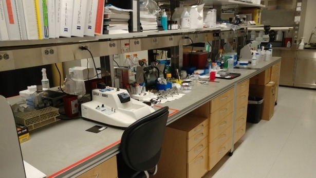
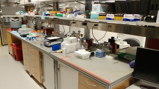
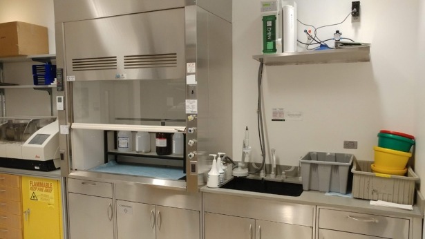
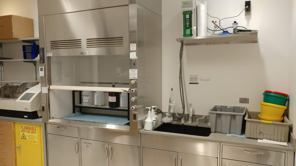
Sample Preparation Lab
We have multiple lab benches for simple sample preparation work including a supply of various materials and disposables. Outside chemicals can be stored in the designated cabinets. Please exercise appropriate lab safety, i.e., disposal of used chemicals and waste container replacement. A fume hood, Milli-Q water purification system, refrigerators (20°C), centrifuge, and hybridization oven are also available on site.


Supplementary Microscope Objectives
We provide a list of the typical objectives installed on our Zeiss fluorescence microscopes along with their corresponding resolutions (lateral and axial) for conventional widefield and confocal microscopy. Please refer to the following inventory for all of the objectives available at the facility. DIC sliders are available for most objectives.
Manufacturer brochures for objective types can also be viewed: Zeiss Objectives, Nikon Objectives, and Olympus Objectives.
How to write Materials & Methods sections and Acknowledgements
In order for other researchers to understand and replicate your findings, it is necessary to thoroughly document both your experimental setup and how the data was acquired. There are many aspects to relate including basic microscope equipment information (see equipment) in addition to some of the criteria listed below (adapted from NIC at HMS). Please ask us if you are unsure or have any questions.
- Make and model of microscope (e.g., Zeiss LSM 980)
- Description of advanced imaging modality – confocal, TIRF, super-resolution, etc. (e.g., Zeiss Airyscan)
- Hardware or software auto-focusing, if used (e.g., Nikon perfect focus system)
- Type, magnification, and numerical aperture of objective lenses (e.g., Zeiss Plan-Apochromat 63x/1.4 Oil)
- Imaging environmental conditions – chamber, cell culture (for live samples) or mounting media, temperature, buffers, etc.
- List of fluorophores.
- Fluorescent filters, including peak transmission and bandwidth (e.g., Chroma 490/30 excitation filter and 525/50 emission filter)
- Laser type, line, and selection method used (e.g., 488nm line from an argon laser, selected with a 488/10nm filter versus 488nm line from an argon laser, selected with an AOTF)
- Make and model of camera (e.g., Hamamatsu ORCA-Flash4.0 V3 sCMOS camera)
- Other motorized components (e.g., Märzhäuser Wetzlar 130x100 STEP linear encoded stage for tile scans)
- Acquisition software make and version (e.g., ZEN Black 2.3 SP1)
- Image analysis software and programming tools detailing any image processing algorithms employed
It is best to document each microscope component (e.g., camera, objective lens) used during your experiment. You are encouraged to send a draft of your Materials & Methods section to us – we will be happy to edit to ensure accuracy. Lastly, if you collected images at the facility, all users should cite Integrative Imaging accordingly in the Acknowledgments section of your manuscript. When staff has helped beyond basic training and provided valuable input, it may also be appropriate to include authorship.
Here are examples of a complete Materials & Methods section and Acknowledgments: