Multi-Color Three Dimensional Imaging Using Multifocus Microscopy
Multi-Color 3-Dimensional Imaging Using Multi-Focus Microscopy
This technology advances imaging techniques of biomicroscopy using an innovative type of a wide-field Multi-Focus microscope to enable fast, high-resolution 3D imaging.
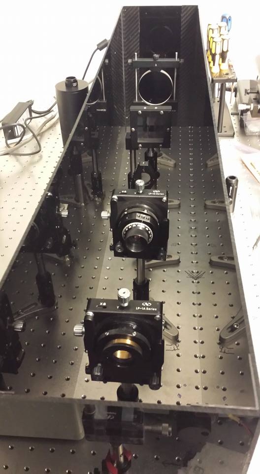
Today, many three-dimensional (3D) biological specimens and processes are studied in two-dimensions (2D) due to the slow speed of the light microscope in recording a 3D image. This technology advances imaging techniques of biomicroscopy using an innovative type of a wide-field Multi-Focus microscope to enable fast, high-resolution 3D imaging. The system includes a novel grating design and chromatic correction scheme that is appended to the camera port of a high-resolution epifluorescence microscope to produce an instant focal stack of high-resolution 2D images simultaneously displayed on a single camera. In addition, the 3D microscope is designed to minimize photobleaching and phototoxicity while enabling high-speed imaging of weak fluorescent samples such as single fluorophores and to acquire multiple focal planes without aberrations to avoid loss in resolution and contrast.
The concept of Multi-Focus imaging uses optical manipulation to produce an entire focal series instantaneously. The Multi-Focus system developed is based on a binary phase plate with a specially designed pattern placed in the Fourier plane, a plane with special optical properties, which allows the grating to split up the emission light from the sample into a set of diffraction orders. Each diffraction order forms a separate image of the sample. The resulting series of images is recorded as a 2D array on a single, large-chip electron-multiplying charge-coupled device camera.
Publications:
The publications and supplemental materials contain guides to recreate the MFM setup.
- Fast multicolor 3D imaging using aberration-corrected multifocus microscopy Nature Methods. 2013;10(1):60-3. https://doi.org/10.1038/nmeth.2277
- MultiFocus Polarization Microscope (MF-PolScope) for 3D polarization imaging of up to 25 focal planes simultaneously Optics Express Vol. 23, Issue 6, pp. 7734-7754 (2015). https://doi.org/10.1364/OE.23.007734
- Multifocus microscopy with precise color multi-phase diffractive optics applied in functional neuronal imaging Biomedical Optics Express. 2016 Feb 16;7(3):855-69. https://doi.org/10.1364/BOE.7.000855
Advantages:
- A powerful tool for fast and sensitive fluorescence microscopy of live samples
- The light efficiency of the imaging path is better than 50 percent
- The current design allows imaging of a volume of 33x33x18 microns at 31Hz
Applications:
- Basic life science research
- Fluorescence biomicroscopy
- Single-particle tracking in live cells
- Neuronal imaging
- Development imaging of small organisms
News:
Microscope expert develops powerful new tools for biologists - 2019
Issued Patent:
United States 9,477,091
How to get it:
- Free for Non-Profit Research. Please contact Innovation Management at innovation@janelia.hhmi.org.
- Commercial Licenses available (Commercial Licensing Managed by David Fung, UCSF)
For inquiries, please reference:
Janelia 2012-006

