Filter
Associated Lab
- Betzig Lab (2) Apply Betzig Lab filter
- Chklovskii Lab (1) Apply Chklovskii Lab filter
- Cui Lab (1) Apply Cui Lab filter
- Druckmann Lab (2) Apply Druckmann Lab filter
- Harris Lab (2) Apply Harris Lab filter
- Ji Lab (2) Apply Ji Lab filter
- Remove Magee Lab filter Magee Lab
- Podgorski Lab (1) Apply Podgorski Lab filter
- Romani Lab (2) Apply Romani Lab filter
- Spruston Lab (2) Apply Spruston Lab filter
- Svoboda Lab (1) Apply Svoboda Lab filter
Associated Support Team
Publication Date
31 Janelia Publications
Showing 11-20 of 31 resultsThe apical tuft is the most remote area of the dendritic tree of neocortical pyramidal neurons. Despite its distal location, the apical dendritic tuft of layer 5 pyramidal neurons receives substantial excitatory synaptic drive and actively processes corticocortical input during behavior. The properties of the voltage-activated ion channels that regulate synaptic integration in tuft dendrites have, however, not been thoroughly investigated. Here, we use electrophysiological and optical approaches to examine the subcellular distribution and function of hyperpolarization-activated cyclic nucleotide-gated nonselective cation (HCN) channels in rat layer 5B pyramidal neurons. Outside-out patch recordings demonstrated that the amplitude and properties of ensemble HCN channel activity were uniform in patches excised from distal apical dendritic trunk and tuft sites. Simultaneous apical dendritic tuft and trunk whole-cell current-clamp recordings revealed that the pharmacological blockade of HCN channels decreased voltage compartmentalization and enhanced the generation and spread of apical dendritic tuft and trunk regenerative activity. Furthermore, multisite two-photon glutamate uncaging demonstrated that HCN channels control the amplitude and duration of synaptically evoked regenerative activity in the distal apical dendritic tuft. In contrast, at proximal apical dendritic trunk and somatic recording sites, the blockade of HCN channels decreased excitability. Dynamic-clamp experiments revealed that these compartment-specific actions of HCN channels were heavily influenced by the local and distributed impact of the high density of HCN channels in the distal apical dendritic arbor. The properties and subcellular distribution pattern of HCN channels are therefore tuned to regulate the interaction between integration compartments in layer 5B pyramidal neurons.
The excitability of individual dendritic branches is a plastic property of neurons. We found that experience in an enriched environment increased propagation of dendritic Na(+) spikes in a subset of individual dendritic branches in rat hippocampal CA1 pyramidal neurons and that this effect was mainly mediated by localized downregulation of A-type K(+) channel function. Thus, dendritic plasticity might be used to store recent experience in individual branches of the dendritic arbor.
Cortical information processing is under state-dependent control of subcortical neuromodulatory systems. Although this modulatory effect is thought to be mediated mainly by slow nonsynaptic metabotropic receptors, other mechanisms, such as direct synaptic transmission, are possible. Yet, it is currently unknown if any such form of subcortical control exists. Here, we present direct evidence of a strong, spatiotemporally precise excitatory input from an ascending neuromodulatory center. Selective stimulation of serotonergic median raphe neurons produced a rapid activation of hippocampal interneurons. At the network level, this subcortical drive was manifested as a pattern of effective disynaptic GABAergic inhibition that spread throughout the circuit. This form of subcortical network regulation should be incorporated into current concepts of normal and pathological cortical function.
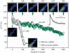
Pulsed lasers are key elements in nonlinear bioimaging techniques such as two-photon fluorescence excitation (TPE) microscopy. Typically, however, only a percent or less of the laser power available can be delivered to the sample before photoinduced damage becomes excessive. Here we describe a passive pulse splitter that converts each laser pulse into a fixed number of sub-pulses of equal energy. We applied the splitter to TPE imaging of fixed mouse brain slices labeled with GFP and show that, in different power regimes, the splitter can be used either to increase the signal rate more than 100-fold or to reduce the rate of photobleaching by over fourfold. In living specimens, the gains were even greater: a ninefold reduction in photobleaching during in vivo imaging of Caenorhabditis elegans larvae, and a six- to 20-fold decrease in the rate of photodamage during calcium imaging of rat hippocampal brain slices.
Pulsed lasers are key elements in nonlinear bioimaging techniques such as two-photon fluorescence excitation (TPE) microscopy. Typically, however, only a percent or less of the laser power available can be delivered to the sample before photoinduced damage becomes excessive. Here we describe a passive pulse splitter that converts each laser pulse into a fixed number of sub-pulses of equal energy. We applied the splitter to TPE imaging of fixed mouse brain slices labeled with GFP and show that, in different power regimes, the splitter can be used either to increase the signal rate more than 100-fold or to reduce the rate of photobleaching by over fourfold. In living specimens, the gains were even greater: a ninefold reduction in photobleaching during in vivo imaging of Caenorhabditis elegans larvae, and a six- to 20-fold decrease in the rate of photodamage during calcium imaging of rat hippocampal brain slices.
Commentary: Na Ji came to me early in her postdoc with an idea to reduce photodamage in nonlinear microscopy by splitting the pulses from an ultrafast laser into multiple subpulses of reduced energy. In six weeks, we constructed a prototype pulse splitter and obtained initial results confirming the validity of her vision. Further experiments with Jeff Magee demonstrated that the splitter could be used to increase imaging speed or reduce photodamage in two photon microscopy by one to two orders of magnitude. This project is a great example of how quickly one can react and exploit new ideas in the Janelia environment.
Phase precession is a well known phenomenon in which a hippocampal place cell will fire action potentials at successively earlier phases (relative to the theta-band oscillations recorded in the local field potential) as an animal moves through the cell’s receptive field (also known as a place field). We present a model in which CA1 pyramidal cell spiking is driven by dual input components arising from CA3 and EC3. The receptive fields of these two input components overlap but are offset in space from each other such that as the animal moves through the model place field, action potentials are driven first by the CA3 input component and then the EC3 input component. As CA3 synaptic input is known to arrive in CA1 at a later theta phase than EC3 input (Mizuseki et al., 2009; Montgomery et al., 2009), CA1 spiking advances in phase as the model transitions from CA3-driven spiking to EC3-driven spiking. Here spike phase is a function of animal location, placing our results in agreement with many experimental observations characterizing CA1 phase precession (O’Keefe and Recce, 1993; Huxter et al., 2003; Geisler et al., 2007). We predict that experimental manipulations that dramatically enhance or disrupt activity in either of these areas should have a significant effect on phase precession observed in CA1.
Understanding the neural correlates of behavior in the mammalian cortex requires measurements of activity in awake, behaving animals. Rodents have emerged as a powerful model for dissecting the cortical circuits underlying behavior attributable to the convergence of several methods. Genetically encoded calcium indicators combined with viral-mediated or transgenic tools enable chronic monitoring of calcium signals in neuronal populations and subcellular structures of identified cell types. Stable one- and two-photon imaging of neuronal activity in awake, behaving animals is now possible using new behavioral paradigms in head-fixed animals, or using novel miniature head-mounted microscopes in freely moving animals. This mini-symposium will highlight recent applications of these methods for studying sensorimotor integration, decision making, learning, and memory in cortical and subcortical brain areas. We will outline future prospects and challenges for identifying the neural underpinnings of task-dependent behavior using cellular imaging in rodents.
Spatial and temporal features of synaptic inputs engage integration mechanisms on multiple scales, including presynaptic release sites, postsynaptic dendrites, and networks of inhibitory interneurons. Here we investigate how these mechanisms cooperate to filter synaptic input in hippocampal area CA1. Dendritic recordings from CA1 pyramidal neurons reveal that proximal inputs from CA3 as well as distal inputs from entorhinal cortex layer III (ECIII) sum sublinearly or linearly at low firing rates due to feedforward inhibition, but sum supralinearly at high firing rates due to synaptic facilitation, producing a high-pass filter. However, during ECIII and CA3 input comparison, supralinear dendritic integration is dynamically balanced by feedforward and feedback inhibition, resulting in suppression of dendritic complex spiking. We find that a particular subpopulation of CA1 interneurons expressing neuropeptide Y (NPY) contributes prominently to this dynamic filter by integrating both ECIII and CA3 input pathways and potently inhibiting CA1 pyramidal neuron dendrites.
Place cells in the CA1 region of the hippocampus express location-specific firing despite receiving a steady barrage of heterogeneously tuned excitatory inputs that should compromise output dynamic range and timing. We examined the role of synaptic inhibition in countering the deleterious effects of off-target excitation. Intracellular recordings in behaving mice demonstrate that bimodal excitation drives place cells, while unimodal excitation drives weaker or no spatial tuning in interneurons. Optogenetic hyperpolarization of interneurons had spatially uniform effects on place cell membrane potential dynamics, substantially reducing spatial selectivity. These data and a computational model suggest that spatially uniform inhibitory conductance enhances rate coding in place cells by suppressing out-of-field excitation and by limiting dendritic amplification. Similarly, we observed that inhibitory suppression of phasic noise generated by out-of-field excitation enhances temporal coding by expanding the range of theta phase precession. Thus, spatially uniform inhibition allows proficient and flexible coding in hippocampal CA1 by suppressing heterogeneously tuned excitation.
Dendritic ion channels play a critical role in shaping synaptic input and are fundamentally important for synaptic integration and plasticity. In the hippocampal region CA1, somato-dendritic gradients of AMPA receptors and the hyperpolarization-activated cation conductance (I(h)) counteract the effects of dendritic filtering on the amplitude, time-course, and temporal integration of distal Schaffer collateral (SC) synaptic inputs within stratum radiatum (SR). While ion channel gradients in CA1 distal apical trunk dendrites within SR have been well characterized, little is known about the patterns of ion channel expression in the distal apical tuft dendrites within stratum lacunosum moleculare (SLM) that receive distinct input from the entorhinal cortex via perforant path (PP) axons. Here, we measured local ion channels densities within these distal apical tuft dendrites to determine if the somato-dendritic gradients of I(h) and AMPA receptors extend into distal tuft dendrites. We also determined the densities of voltage-gated sodium channels and NMDA receptors. We found that the densities of AMPA receptors, I(h,) and voltage-gated sodium channels are similar in tuft dendrites in SLM when compared with distal apical dendrites in SR, while the ratio of NMDA receptors to AMPA receptors increases in tuft dendrites relative to distal apical dendrites within SR. These data indicate that the somato-dendritic gradients of I(h) and AMPA receptors in apical dendrites do not extend into the distal tuft, and the relative densities of voltage-gated sodium channels and NMDA receptors are poised to support nonlinear integration of correlated SC and PP input.