Filter
Publication Date
- Remove October 1, 2015 filter October 1, 2015
- Remove October 2015 filter October 2015
- Remove 2015 filter 2015
2 Janelia Publications
Showing 1-2 of 2 results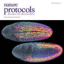
Light-sheet microscopy is a powerful method for imaging the development and function of complex biological systems at high spatiotemporal resolution and over long time scales. Such experiments typically generate terabytes of multidimensional image data, and thus they demand efficient computational solutions for data management, processing and analysis. We present protocols and software to tackle these steps, focusing on the imaging-based study of animal development. Our protocols facilitate (i) high-speed lossless data compression and content-based multiview image fusion optimized for multicore CPU architectures, reducing image data size 30–500-fold; (ii) automated large-scale cell tracking and segmentation; and (iii) visualization, editing and annotation of multiterabyte image data and cell-lineage reconstructions with tens of millions of data points. These software modules are open source. They provide high data throughput using a single computer workstation and are readily applicable to a wide spectrum of biological model systems.