Filter
Associated Lab
- Ahrens Lab (3) Apply Ahrens Lab filter
- Aso Lab (6) Apply Aso Lab filter
- Baker Lab (2) Apply Baker Lab filter
- Betzig Lab (11) Apply Betzig Lab filter
- Branson Lab (6) Apply Branson Lab filter
- Cardona Lab (5) Apply Cardona Lab filter
- Chklovskii Lab (2) Apply Chklovskii Lab filter
- Cui Lab (5) Apply Cui Lab filter
- Dickson Lab (1) Apply Dickson Lab filter
- Druckmann Lab (3) Apply Druckmann Lab filter
- Dudman Lab (3) Apply Dudman Lab filter
- Eddy/Rivas Lab (4) Apply Eddy/Rivas Lab filter
- Egnor Lab (1) Apply Egnor Lab filter
- Fetter Lab (5) Apply Fetter Lab filter
- Freeman Lab (7) Apply Freeman Lab filter
- Funke Lab (1) Apply Funke Lab filter
- Gonen Lab (5) Apply Gonen Lab filter
- Grigorieff Lab (7) Apply Grigorieff Lab filter
- Harris Lab (7) Apply Harris Lab filter
- Heberlein Lab (1) Apply Heberlein Lab filter
- Hess Lab (7) Apply Hess Lab filter
- Jayaraman Lab (4) Apply Jayaraman Lab filter
- Ji Lab (4) Apply Ji Lab filter
- Karpova Lab (1) Apply Karpova Lab filter
- Keleman Lab (2) Apply Keleman Lab filter
- Keller Lab (7) Apply Keller Lab filter
- Lavis Lab (5) Apply Lavis Lab filter
- Leonardo Lab (2) Apply Leonardo Lab filter
- Liu (Zhe) Lab (4) Apply Liu (Zhe) Lab filter
- Looger Lab (9) Apply Looger Lab filter
- Magee Lab (5) Apply Magee Lab filter
- Murphy Lab (1) Apply Murphy Lab filter
- Pastalkova Lab (3) Apply Pastalkova Lab filter
- Reiser Lab (2) Apply Reiser Lab filter
- Romani Lab (2) Apply Romani Lab filter
- Rubin Lab (16) Apply Rubin Lab filter
- Saalfeld Lab (3) Apply Saalfeld Lab filter
- Scheffer Lab (2) Apply Scheffer Lab filter
- Schreiter Lab (4) Apply Schreiter Lab filter
- Shroff Lab (1) Apply Shroff Lab filter
- Simpson Lab (4) Apply Simpson Lab filter
- Singer Lab (10) Apply Singer Lab filter
- Spruston Lab (7) Apply Spruston Lab filter
- Stern Lab (4) Apply Stern Lab filter
- Sternson Lab (7) Apply Sternson Lab filter
- Svoboda Lab (9) Apply Svoboda Lab filter
- Tjian Lab (6) Apply Tjian Lab filter
- Truman Lab (6) Apply Truman Lab filter
- Turaga Lab (1) Apply Turaga Lab filter
- Turner Lab (2) Apply Turner Lab filter
- Wu Lab (1) Apply Wu Lab filter
- Zlatic Lab (4) Apply Zlatic Lab filter
- Zuker Lab (2) Apply Zuker Lab filter
Associated Project Team
Associated Support Team
- Electron Microscopy (1) Apply Electron Microscopy filter
- Gene Targeting and Transgenics (3) Apply Gene Targeting and Transgenics filter
- Invertebrate Shared Resource (1) Apply Invertebrate Shared Resource filter
- Janelia Experimental Technology (2) Apply Janelia Experimental Technology filter
- Management Team (1) Apply Management Team filter
- Molecular Genomics (1) Apply Molecular Genomics filter
- Primary & iPS Cell Culture (2) Apply Primary & iPS Cell Culture filter
- Project Technical Resources (1) Apply Project Technical Resources filter
- Scientific Computing Software (11) Apply Scientific Computing Software filter
Publication Date
- December 2015 (15) Apply December 2015 filter
- November 2015 (22) Apply November 2015 filter
- October 2015 (16) Apply October 2015 filter
- September 2015 (16) Apply September 2015 filter
- August 2015 (17) Apply August 2015 filter
- July 2015 (18) Apply July 2015 filter
- June 2015 (16) Apply June 2015 filter
- May 2015 (16) Apply May 2015 filter
- April 2015 (18) Apply April 2015 filter
- March 2015 (16) Apply March 2015 filter
- February 2015 (15) Apply February 2015 filter
- January 2015 (10) Apply January 2015 filter
- Remove 2015 filter 2015
195 Janelia Publications
Showing 181-190 of 195 resultsUNLABELLED: Sensorimotor delays decouple behaviors from the events that drive them. The brain compensates for these delays with predictive mechanisms, but the efficacy and timescale over which these mechanisms operate remain poorly understood. Here, we assess how prediction is used to compensate for prey movement that occurs during visuomotor processing. We obtained high-speed video records of freely moving, tongue-projecting salamanders catching walking prey, emulating natural foraging conditions. We found that tongue projections were preceded by a rapid head turn lasting ∼130 ms. This motor lag, combined with the ∼100 ms phototransduction delay at photopic light levels, gave a ∼230 ms visuomotor response delay during which prey typically moved approximately one body length. Tongue projections, however, did not significantly lag prey position but were highly accurate instead. Angular errors in tongue projection accuracy were consistent with a linear extrapolation model that predicted prey position at the time of tongue contact using the average prey motion during a ∼175 ms period one visual latency before the head movement. The model explained successful strikes where the tongue hit the fly, and unsuccessful strikes where the fly turned and the tongue hit a phantom location consistent with the fly's earlier trajectory. The model parameters, obtained from the data, agree with the temporal integration and latency of retinal responses proposed to contribute to motion extrapolation. These results show that the salamander predicts future prey position and that prediction significantly improves prey capture success over a broad range of prey speeds and light levels. SIGNIFICANCE STATEMENT: Neural processing delays cause actions to lag behind the events that elicit them. To cope with these delays, the brain predicts what will happen in the future. While neural circuits in the retina and beyond have been suggested to participate in such predictions, few behaviors have been explored sufficiently to constrain circuit function. Here we show that salamanders aim their tongues by using extrapolation to estimate future prey position, thereby compensating for internal delays from both visual and motor processing. Predictions made just before a prey turn resulted in the tongue being projected to a position consistent with the prey's pre-turn trajectory. These results define the computations and operating regimen for neural circuits that predict target motion.
To facilitate large scale functional studies in Drosophila, the Drosophila Transgenic RNAi Project (TRiP) at Harvard Medical School (HMS) was established along with several goals: developing efficient vectors for RNAi that work in all tissues, generating a genome scale collection of RNAi stocks with input from the community, distributing the lines as they are generated through existing stock centers, validating as many lines as possible using RT-qPCR and phenotypic analyses, and developing tools and web resources for identifying RNAi lines and retrieving existing information on their quality. With these goals in mind, here we describe in detail the various tools we developed and the status of the collection, which is currently comprised of 11,491 lines and covering 71% of Drosophila genes. Data on the characterization of the lines either by RT-qPCR or phenotype is available on a dedicated web site, the RNAi Stock Validation and Phenotypes Project (RSVP; www.flyrnai.org/RSVP.html), and stocks are available from three stock centers, the Bloomington Drosophila Stock Center (USA), National Institute of Genetics (Japan), and TsingHua Fly Center (China).
Sensory cue inputs and memory-related internal brain activities govern the firing of hippocampal neurons, but which specific firing patterns are induced by either of the two processes remains unclear. We found that sensory cues guided the firing of neurons in rats on a timescale of seconds and supported the formation of spatial firing fields. Independently of the sensory inputs, the memory-related network activity coordinated the firing of neurons not only on a second-long timescale, but also on a millisecond-long timescale, and was dependent on medial septum inputs. We propose a network mechanism that might coordinate this internally generated firing. Overall, we suggest that two independent mechanisms support the formation of spatial firing fields in hippocampus, but only the internally organized system supports short-timescale sequential firing and episodic memory.
Using a combination of metabolically labeled glycans, a bioorthogonal copper(I)-catalyzed azide-alkyne cycloaddition, and the controlled bleaching of fluorescent probes conjugated to azide- or alkyne-tagged glycans, a sufficiently low spatial density of dye-labeled glycans was achieved, enabling dynamic single-molecule tracking and super-resolution imaging of N-linked sialic acids and O-linked N-acetyl galactosamine (GalNAc) on the membrane of live cells. Analysis of the trajectories of these dye-labeled glycans in mammary cancer cells revealed constrained diffusion of both N- and O-linked glycans, which was interpreted as reflecting the mobility of the glycan rather than to be caused by transient immobilization owing to spatial inhomogeneities on the plasma membrane. Stochastic optical reconstruction microscopy (STORM) imaging revealed the structure of dynamic membrane nanotubes.
Analysis of single molecules in living cells has provided quantitative insights into the kinetics of fundamental biological processes; however, the dynamics of messenger RNA (mRNA) translation have yet to be addressed. We have developed a fluorescence microscopy technique that reports on the first translation events of individual mRNA molecules. This allowed us to examine the spatiotemporal regulation of translation during normal growth and stress and during Drosophila oocyte development. We have shown that mRNAs are not translated in the nucleus but translate within minutes after export, that sequestration within P-bodies regulates translation, and that oskar mRNA is not translated until it reaches the posterior pole of the oocyte. This methodology provides a framework for studying initiation of protein synthesis on single mRNAs in living cells.
Focused-ion-beam scanning electron microscopy (FIB-SEM) has become an essential tool for studying neural tissue at resolutions below 10 nm × 10 nm × 10 nm, producing data sets optimized for automatic connectome tracing. We present a technical advance, ultrathick sectioning, which reliably subdivides embedded tissue samples into chunks (20 μm thick) optimally sized and mounted for efficient, parallel FIB-SEM imaging. These chunks are imaged separately and then 'volume stitched' back together, producing a final three-dimensional data set suitable for connectome tracing.
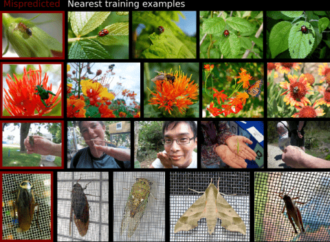
Modern supervised learning algorithms can learn very accurate and complex discriminating functions. But when these classifiers fail, this complexity can also be a drawback because there is no easy, intuitive way to diagnose why they are failing and remedy the problem. This important question has received little attention. To address this problem, we propose a novel method to analyze and understand a classifier's errors. Our method centers around a measure of how much influence a training example has on the classifier's prediction for a test example. To understand why a classifier is mispredicting the label of a given test example, the user can find and review the most influential training examples that caused this misprediction, allowing them to focus their attention on relevant areas of the data space. This will aid the user in determining if and how the training data is inconsistently labeled or lacking in diversity, or if the feature representation is insufficient. As computing the influence of each training example is computationally impractical, we propose a novel distance metric to approximate influence for boosting classifiers that is fast enough to be used interactively. We also show several novel use paradigms of our distance metric. Through experiments, we show that it can be used to find incorrectly or inconsistently labeled training examples, to find specific areas of the data space that need more training data, and to gain insight into which features are missing from the current representation. Code is available at https://github.com/kristinbranson/InfluentialNeighbors.
Vinculin is filamentous (F)-actin-binding protein enriched in integrin-based adhesions to the extracellular matrix (ECM). Whereas studies in 2-dimensional (2D) tissue culture models have suggested that vinculin negatively regulates cell migration by promoting cytoskeleton-ECM coupling to strengthen and stabilize adhesions, its role in regulating cell migration in more physiologic, 3-dimensional (3D) environments is unclear. To address the role of vinculin in 3D cell migration, we analyzed the morphodynamics, migration, and ECM remodeling of primary murine embryonic fibroblasts (MEFs) with cre/loxP-mediated vinculin gene disruption in 3D collagen I cultures. We found that vinculin promoted 3D cell migration by increasing directional persistence. Vinculin was necessary for persistent cell protrusion, cell elongation, and stable cell orientation in 3D collagen, but was dispensable for lamellipodia formation, suggesting that vinculin-mediated cell adhesion to the ECM is needed to convert actin-based cell protrusion into persistent cell shape change and migration. Consistent with this finding, vinculin was necessary for efficient traction force generation in 3D collagen without affecting myosin II activity and promoted 3D collagen fiber alignment and macroscopical gel contraction. Our results suggest that vinculin promotes directionally persistent cell migration and tension-dependent ECM remodeling in complex 3D environments by increasing cell-ECM adhesion and traction force generation.-Thievessen, I., Fakhri, N., Steinwachs, J., Kraus, V., McIsaac, R. S., Gao, L., Chen, B.-C., Baird, M. A., Davidson, M. W., Betzig, E., Oldenbourg, R., Waterman, C., M., Fabry, B. Vinculin is required for cell polarization, migration, and extracellular matrix remodeling in 3D collagen.
The simultaneous visualization, identification and targeting of neurons during patch clamp-mediated electrophysiological recordings is a basic technique in neuroscience, yet it is often complicated by the inability to visualize the pipette tip, particularly in deep brain tissue. Here we demonstrate a novel approach in which fluorescent quantum dot probes are used to coat pipettes prior to their use. The strong two-photon absorption cross sections of the quantum dots afford robust contrast at significantly deeper penetration depths than current methods allow. We demonstrate the utility of this technique in multiple recording formats both in vitro and in vivo where imaging of the pipettes is achieved at remarkable depths (up to 800 microns). Notably, minimal perturbation of cellular physiology is observed over the hours-long time course of neuronal recordings. We discuss our results within the context of the role that quantum dot nanoprobes may play in understanding neuronal cell physiology.