Filter
Associated Lab
- Baker Lab (2) Apply Baker Lab filter
- Betzig Lab (3) Apply Betzig Lab filter
- Cardona Lab (2) Apply Cardona Lab filter
- Chklovskii Lab (1) Apply Chklovskii Lab filter
- Druckmann Lab (1) Apply Druckmann Lab filter
- Eddy/Rivas Lab (3) Apply Eddy/Rivas Lab filter
- Fetter Lab (2) Apply Fetter Lab filter
- Harris Lab (1) Apply Harris Lab filter
- Hess Lab (2) Apply Hess Lab filter
- Jayaraman Lab (2) Apply Jayaraman Lab filter
- Ji Lab (1) Apply Ji Lab filter
- Keller Lab (2) Apply Keller Lab filter
- Looger Lab (6) Apply Looger Lab filter
- Magee Lab (3) Apply Magee Lab filter
- Reiser Lab (2) Apply Reiser Lab filter
- Riddiford Lab (2) Apply Riddiford Lab filter
- Rubin Lab (2) Apply Rubin Lab filter
- Saalfeld Lab (1) Apply Saalfeld Lab filter
- Scheffer Lab (3) Apply Scheffer Lab filter
- Shroff Lab (1) Apply Shroff Lab filter
- Simpson Lab (3) Apply Simpson Lab filter
- Sternson Lab (1) Apply Sternson Lab filter
- Svoboda Lab (7) Apply Svoboda Lab filter
- Truman Lab (4) Apply Truman Lab filter
Associated Project Team
Associated Support Team
Publication Date
- December 2010 (3) Apply December 2010 filter
- November 2010 (4) Apply November 2010 filter
- October 2010 (5) Apply October 2010 filter
- September 2010 (3) Apply September 2010 filter
- August 2010 (7) Apply August 2010 filter
- July 2010 (2) Apply July 2010 filter
- June 2010 (6) Apply June 2010 filter
- May 2010 (3) Apply May 2010 filter
- April 2010 (5) Apply April 2010 filter
- March 2010 (1) Apply March 2010 filter
- February 2010 (6) Apply February 2010 filter
- January 2010 (16) Apply January 2010 filter
- Remove 2010 filter 2010
61 Janelia Publications
Showing 31-40 of 61 resultsThe role of gamma amino butyric acid (GABA) release and inhibitory neurotransmission in regulating most behaviors remains unclear. The vesicular GABA transporter (VGAT) is required for the storage of GABA in synaptic vesicles and provides a potentially useful probe for inhibitory circuits. However, specific pharmacologic agents for VGAT are not available, and VGAT knockout mice are embryonically lethal, thus precluding behavioral studies. We have identified the Drosophila ortholog of the vesicular GABA transporter gene (which we refer to as dVGAT), immunocytologically mapped dVGAT protein expression in the larva and adult and characterized a dVGAT(minos) mutant allele. dVGAT is embryonically lethal and we do not detect residual dVGAT expression, suggesting that it is either a strong hypomorph or a null. To investigate the function of VGAT and GABA signaling in adult visual flight behavior, we have selectively rescued the dVGAT mutant during development. We show that reduced GABA release does not compromise the active optomotor control of wide-field pattern motion. Conversely, reduced dVGAT expression disrupts normal object tracking and figure-ground discrimination. These results demonstrate that visual behaviors are segregated by the level of GABA signaling in flies, and more generally establish dVGAT as a model to study the contribution of GABA release to other complex behaviors.
Cell adhesions to the extracellular matrix (ECM) are necessary for morphogenesis, immunity, and wound healing. Focal adhesions are multifunctional organelles that mediate cell-ECM adhesion, force transmission, cytoskeletal regulation and signaling. Focal adhesions consist of a complex network of trans-plasma-membrane integrins and cytoplasmic proteins that form a <200-nm plaque linking the ECM to the actin cytoskeleton. The complexity of focal adhesion composition and dynamics implicate an intricate molecular machine. However, focal adhesion molecular architecture remains unknown. Here we used three-dimensional super-resolution fluorescence microscopy (interferometric photoactivated localization microscopy) to map nanoscale protein organization in focal adhesions. Our results reveal that integrins and actin are vertically separated by a \~{}40-nm focal adhesion core region consisting of multiple protein-specific strata: a membrane-apposed integrin signaling layer containing integrin cytoplasmic tails, focal adhesion kinase, and paxillin; an intermediate force-transduction layer containing talin and vinculin; and an uppermost actin-regulatory layer containing zyxin, vasodilator-stimulated phosphoprotein and α-actinin. By localizing amino- and carboxy-terminally tagged talins, we reveal talin’s polarized orientation, indicative of a role in organizing the focal adhesion strata. The composite multilaminar protein architecture provides a molecular blueprint for understanding focal adhesion functions.
Imaging approaches based on single molecule localization break the diffraction barrier of conventional fluorescence microscopy, allowing for bioimaging with nanometer resolution. It remains a challenge, however, to precisely localize photon-limited single molecules in 3D. We have developed a new localization-based imaging technique achieving almost isotropic subdiffraction resolution in 3D. A tilted mirror is used to generate a side view in addition to the front view of activated single emitters, allowing their 3D localization to be precisely determined for superresolution imaging. Because both front and side views are in focus, this method is able to efficiently collect emitted photons. The technique is simple to implement on a commercial fluorescence microscope, and especially suitable for biological samples with photon-limited chromophores such as endogenously expressed photoactivatable fluorescent proteins. Moreover, this method is relatively resistant to optical aberration, as it requires only centroid determination for localization analysis. Here we demonstrate the application of this method to 3D imaging of bacterial protein distribution and neuron dendritic morphology with subdiffraction resolution.
Although hippocampal theta oscillations represent a prime example of temporal coding in the mammalian brain, little is known about the specific biophysical mechanisms. Intracellular recordings support a particular abstract oscillatory interference model of hippocampal theta activity, the soma-dendrite interference model. To gain insight into the cellular and circuit level mechanisms of theta activity, we implemented a similar form of interference using the actual hippocampal network in mice in vitro. We found that pairing increasing levels of phasic dendritic excitation with phasic stimulation of perisomatic projecting inhibitory interneurons induced a somatic polarization and action potential timing profile that reproduced most common features. Alterations in the temporal profile of inhibition were required to fully capture all features. These data suggest that theta-related place cell activity is generated through an interaction between a phasic dendritic excitation and a phasic perisomatic shunting inhibition delivered by interneurons, a subset of which undergo activity-dependent presynaptic modulation.
Classical studies have related the spiking of selected neocortical neurons to behavior, but little is known about activity sampled from the entire neural population. We recorded from neurons selected independent of spiking, using cell-attached recordings and two-photon calcium imaging, in the barrel cortex of mice performing an object localization task. Spike rates varied across neurons, from silence to >60 Hz. Responses were diverse, with some neurons showing large increases in spike rate when whiskers contacted the object. Nearly half the neurons discriminated object location; a small fraction of neurons discriminated perfectly. More active neurons were more discriminative. Layer (L) 4 and L5 contained the highest fractions of discriminating neurons (\~{}63% and 79%, respectively), but a few L2/3 neurons were also highly discriminating. Approximately 13,000 spikes per activated barrel column were available to mice for decision making. Coding of object location in the barrel cortex is therefore highly redundant.
A striking aspect of cortical neural networks is the divergence of a relatively small number of input channels from the peripheral sensory apparatus into a large number of cortical neurons, an over-complete representation strategy. Cortical neurons are then connected by a sparse network of lateral synapses. Here we propose that such architecture may increase the persistence of the representation of an incoming stimulus, or a percept. We demonstrate that for a family of networks in which the receptive field of each neuron is re-expressed by its outgoing connections, a represented percept can remain constant despite changing activity. We term this choice of connectivity REceptive FIeld REcombination (REFIRE) networks. The sparse REFIRE network may serve as a high-dimensional integrator and a biologically plausible model of the local cortical circuit.
Recently it was demonstrated that long-lived quantum coherence exists during excitation energy transport in photosynthesis. It is a valid question up to which length, time and mass scales quantum coherence may extend, how one may detect this coherence and what, if any, role it plays in the dynamics of the system. Here we suggest that the selectivity filter of ion channels may exhibit quantum coherence, which might be relevant for the process of ion selectivity and conduction. We show that quantum resonances could provide an alternative approach to ultrafast two-dimensional (2D) spectroscopy to probe these quantum coherences. We demonstrate that the emergence of resonances in the conduction of ion channels that are modulated periodically by time-dependent external electric fields can serve as signatures of quantum coherence in such a system. Assessments of experimental feasibility and specific paths towards the experimental realization of such experiments are presented.

Novel approaches to bio-imaging and automated computational image processing allow the design of truly quantitative studies in developmental biology. Cell behavior, cell fate decisions, cell interactions during tissue morphogenesis, and gene expression dynamics can be analyzed in vivo for entire complex organisms and throughout embryonic development. We review state-of-the-art technology for live imaging, focusing on fluorescence light microscopy techniques for system-level investigations of animal development and discuss computational approaches to image segmentation, cell tracking, automated data annotation, and biophysical modeling. We argue that the substantial increase in data complexity and size requires sophisticated new strategies to data analysis to exploit the enormous potential of these new resources.
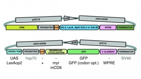
A wide variety of biological experiments rely on the ability to express an exogenous gene in a transgenic animal at a defined level and in a spatially and temporally controlled pattern. We describe major improvements of the methods available for achieving this objective in Drosophila melanogaster. We have systematically varied core promoters, UTRs, operator sequences, and transcriptional activating domains used to direct gene expression with the GAL4, LexA, and Split GAL4 transcription factors and the GAL80 transcriptional repressor. The use of site-specific integration allowed us to make quantitative comparisons between different constructs inserted at the same genomic location. We also characterized a set of PhiC31 integration sites for their ability to support transgene expression of both drivers and responders in the nervous system. The increased strength and reliability of these optimized reagents overcome many of the previous limitations of these methods and will facilitate genetic manipulations of greater complexity and sophistication.
The secondary neurons generated in the thoracic central nervous system of Drosophila arise from a hemisegmental set of 25 neuronal stem cells, the neuroblasts (NBs). Each NB undergoes repeated asymmetric divisions to produce a series of smaller ganglion mother cells (GMCs), which typically divide once to form two daughter neurons. We find that the two daughters of the GMC consistently have distinct fates. Using both loss-of-function and gain-of-function approaches, we examined the role of Notch signaling in establishing neuronal fates within all of the thoracic secondary lineages. In all cases, the ’A’ (Notch(ON)) sibling assumes one fate and the ’B’ (Notch(OFF)) sibling assumes another, and this relationship holds throughout the neurogenic period, resulting in two major neuronal classes: the A and B hemilineages. Apparent monotypic lineages typically result from the death of one sibling throughout the lineage, resulting in a single, surviving hemilineage. Projection neurons are predominantly from the B hemilineages, whereas local interneurons are typically from A hemilineages. Although sibling fate is dependent on Notch signaling, it is not necessarily dependent on numb, a gene classically involved in biasing Notch activation. When Numb was removed at the start of larval neurogenesis, both A and B hemilineages were still generated, but by the start of the third larval instar, the removal of Numb resulted in all neurons assuming the A fate. The need for Numb to direct Notch signaling correlated with a decrease in NB cell cycle time and may be a means for coping with multiple sibling pairs simultaneously undergoing fate decisions.