Filter
Associated Lab
- Betzig Lab (4) Apply Betzig Lab filter
- Fetter Lab (1) Apply Fetter Lab filter
- Gonen Lab (1) Apply Gonen Lab filter
- Harris Lab (1) Apply Harris Lab filter
- Lavis Lab (5) Apply Lavis Lab filter
- Liu (Zhe) Lab (8) Apply Liu (Zhe) Lab filter
- Rubin Lab (1) Apply Rubin Lab filter
- Singer Lab (5) Apply Singer Lab filter
- Remove Tjian Lab filter Tjian Lab
- Wu Lab (1) Apply Wu Lab filter
Associated Project Team
Publication Date
- 2016 (3) Apply 2016 filter
- 2015 (6) Apply 2015 filter
- 2014 (4) Apply 2014 filter
- 2013 (1) Apply 2013 filter
- 2012 (1) Apply 2012 filter
- 2011 (4) Apply 2011 filter
- 2010 (2) Apply 2010 filter
- 2009 (4) Apply 2009 filter
- 2008 (7) Apply 2008 filter
- 2007 (5) Apply 2007 filter
- 2006 (6) Apply 2006 filter
- 2005 (4) Apply 2005 filter
- 2004 (5) Apply 2004 filter
- 2003 (6) Apply 2003 filter
- 2002 (5) Apply 2002 filter
- 1994 (1) Apply 1994 filter
Type of Publication
64 Publications
Showing 1-10 of 64 results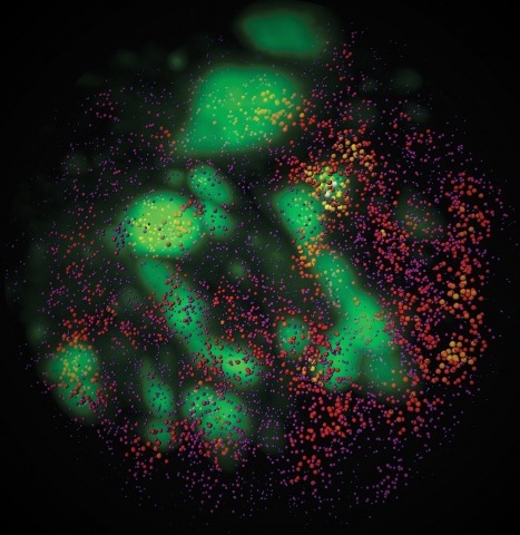
Combinatorial cis-regulatory networks encoded in animal genomes represent the foundational gene expression mechanism for directing cell-fate commitment and maintenance of cell identity by transcription factors (TFs). However, the 3D spatial organization of cis-elements and how such sub-nuclear structures influence TF activity remain poorly understood. Here, we combine lattice light-sheet imaging, single-molecule tracking, numerical simulations, and ChIP-exo mapping to localize and functionally probe Sox2 enhancer-organization in living embryonic stem cells. Sox2 enhancers form 3D-clusters that are segregated from heterochromatin but overlap with a subset of Pol II enriched regions. Sox2 searches for specific binding targets via a 3D-diffusion dominant mode when shuttling long-distances between clusters while chromatin-bound states predominate within individual clusters. Thus, enhancer clustering may reduce global search efficiency but enables rapid local fine-tuning of TF search parameters. Our results suggest an integrated model linking cis-element 3D spatial distribution to local-versus-global target search modalities essential for regulating eukaryotic gene transcription.
Specific labeling of biomolecules with bright fluorophores is the keystone of fluorescence microscopy. Genetically encoded self-labeling tag proteins can be coupled to synthetic dyes inside living cells, resulting in brighter reporters than fluorescent proteins. Intracellular labeling using these techniques requires cell-permeable fluorescent ligands, however, limiting utility to a small number of classic fluorophores. Here we describe a simple structural modification that improves the brightness and photostability of dyes while preserving spectral properties and cell permeability. Inspired by molecular modeling, we replaced the N,N-dimethylamino substituents in tetramethylrhodamine with four-membered azetidine rings. This addition of two carbon atoms doubles the quantum efficiency and improves the photon yield of the dye in applications ranging from in vitro single-molecule measurements to super-resolution imaging. The novel substitution is generalizable, yielding a palette of chemical dyes with improved quantum efficiencies that spans the UV and visible range.
TFIID-a complex of TATA-binding protein (TBP) and TBP-associated factors (TAFs)-is a central component of the Pol II promoter recognition apparatus. Recent studies have revealed significant downregulation of TFIID subunits in terminally differentiated myocytes, hepatocytes and adipocytes. Here, we report that TBP protein levels are tightly regulated by the ubiquitin-proteasome system. Using an in vitro ubiquitination assay coupled with biochemical fractionation, we identified Huwe1 as an E3 ligase targeting TBP for K48-linked ubiquitination and proteasome-mediated degradation. Upregulation of Huwe1 expression during myogenesis induces TBP degradation and myotube differentiation. We found that Huwe1 activity on TBP is antagonized by the deubiquitinase USP10, which protects TBP from degradation. Thus, modulating the levels of both Huwe1 and USP10 appears to fine-tune the requisite degradation of TBP during myogenesis. Together, our study unmasks a previously unknown interplay between an E3 ligase and a deubiquitinating enzyme regulating TBP levels during cellular differentiation.
The sterol regulatory element binding protein (SREBP) family of transcription activators are critical regulators of cholesterol and fatty acid homeostasis. We previously demonstrated that human SREBPs bind the CREB-binding protein (CBP)/p300 acetyltransferase KIX domain and recruit activator-recruited co-factor (ARC)/Mediator co-activator complexes through unknown mechanisms. Here we show that SREBPs use the evolutionarily conserved ARC105 (also called MED15) subunit to activate target genes. Structural analysis of the SREBP-binding domain in ARC105 by NMR revealed a three-helix bundle with marked similarity to the CBP/p300 KIX domain. In contrast to SREBPs, the CREB and c-Myb activators do not bind the ARC105 KIX domain, although they interact with the CBP KIX domain, revealing a surprising specificity among structurally related activator-binding domains. The Caenorhabditis elegans SREBP homologue SBP-1 promotes fatty acid homeostasis by regulating the expression of lipogenic enzymes. We found that, like SBP-1, the C. elegans ARC105 homologue MDT-15 is required for fatty acid homeostasis, and show that both SBP-1 and MDT-15 control transcription of genes governing desaturation of stearic acid to oleic acid. Notably, dietary addition of oleic acid significantly rescued various defects of nematodes targeted with RNA interference against sbp-1 and mdt-15, including impaired intestinal fat storage, infertility, decreased size and slow locomotion, suggesting that regulation of oleic acid levels represents a physiologically critical function of SBP-1 and MDT-15. Taken together, our findings demonstrate that ARC105 is a key effector of SREBP-dependent gene regulation and control of lipid homeostasis in metazoans.
Bromodomains bind acetylated histone H4 peptides in vitro. Since many chromatin remodeling complexes and the general transcription factor TFIID contain bromodomains, they may link histone acetylation to increased transcription. Here we show that yeast Bdf1 bromodomains recognize endogenous acetyl-histone H3/H4 as a mechanism for chromatin association in vivo. Surprisingly, deletion of BDF1 or a Bdf1 mutation that abolishes histone binding leads to transcriptional downregulation of genes located at heterochromatin-euchromatin boundaries. Wild-type Bdf1 protein imposes a physical barrier to the spreading of telomere- and mating-locus-proximal SIR proteins. Biochemical experiments indicate that Bdf1 competes with the Sir2 deacetylase for binding to acetylated histone H4. These data suggest an active role for Bdf1 in euchromatin maintenance and antisilencing through a histone tail-encoded boundary function.
Direct visualization of genomic loci in the 3D nucleus is important for understanding the spatial organization of the genome and its association with gene expression. Various DNA FISH methods have been developed in the past decades, all involving denaturing dsDNA and hybridizing fluorescent nucleic acid probes. Here we report a novel approach that uses in vitro constituted nuclease-deficient clustered regularly interspaced short palindromic repeats (CRISPR)/CRISPR-associated caspase 9 (Cas9) complexes as probes to label sequence-specific genomic loci fluorescently without global DNA denaturation (Cas9-mediated fluorescence in situ hybridization, CASFISH). Using fluorescently labeled nuclease-deficient Cas9 (dCas9) protein assembled with various single-guide RNA (sgRNA), we demonstrated rapid and robust labeling of repetitive DNA elements in pericentromere, centromere, G-rich telomere, and coding gene loci. Assembling dCas9 with an array of sgRNAs tiling arbitrary target loci, we were able to visualize nonrepetitive genomic sequences. The dCas9/sgRNA binary complex is stable and binds its target DNA with high affinity, allowing sequential or simultaneous probing of multiple targets. CASFISH assays using differently colored dCas9/sgRNA complexes allow multicolor labeling of target loci in cells. In addition, the CASFISH assay is remarkably rapid under optimal conditions and is applicable for detection in primary tissue sections. This rapid, robust, less disruptive, and cost-effective technology adds a valuable tool for basic research and genetic diagnosis.
Cell-type-selective expression of the TFIID subunit TAF(II)105 (renamed TAF4b) in the ovary is essential for proper follicle development. Although a multitude of signaling pathways required for folliculogenesis have been identified, downstream transcriptional integrators of these signals remain largely unknown. Here, we show that TAF4b controls the granulosa-cell-specific expression of the proto-oncogene c-jun, and together they regulate transcription of ovary-selective promoters. Instead of using cell-type-specific activators, our findings suggest that the coactivator TAF4b regulates the expression of tissue-specific genes, at least in part, through the cell-type-specific induction of c-jun, a ubiquitous activator. Importantly, the loss of TAF4b in ovarian granulosa cells disrupts cellular morphologies and interactions during follicle growth that likely contribute to the infertility observed in TAF4b-null female mice. These data highlight a mechanism for potentiating tissue-selective functions of the basal transcription machinery and reveal intricate networks of gene expression that orchestrate ovarian-specific functions and cell morphology.

To gain insights into coordinated lineage-specification and morphogenetic processes during early embryogenesis, here we report a systematic identification of transcriptional programs mediated by a key developmental regulator-Brachyury. High-resolution chromosomal localization mapping of Brachyury by ChIP sequencing and ChIP-exonuclease revealed distinct sequence signatures enriched in Brachyury-bound enhancers. A combination of genome-wide in vitro and in vivo perturbation analysis and cross-species evolutionary comparison unveiled a detailed Brachyury-dependent gene-regulatory network that directly links the function of Brachyury to diverse developmental pathways and cellular housekeeping programs. We also show that Brachyury functions primarily as a transcriptional activator genome-wide and that an unexpected gene-regulatory feedback loop consisting of Brachyury, Foxa2, and Sox17 directs proper stem-cell lineage commitment during streak formation. Target gene and mRNA-sequencing correlation analysis of the T(c) mouse model supports a crucial role of Brachyury in up-regulating multiple key hematopoietic and muscle-fate regulators. Our results thus chart a comprehensive map of the Brachyury-mediated gene-regulatory network and how it influences in vivo developmental homeostasis and coordination.
Intrinsically disordered protein regions (IDRs) are peptide segments that fail to form stable 3-dimensional structures in the absence of partner proteins. They are abundant in eukaryotic proteomes and are often associated with human diseases, but their biological functions have been elusive to study. Here we report the identification of a tin(IV) oxochloride-derived cluster that binds an evolutionarily conserved IDR within the metazoan TFIID transcription complex. Binding arrests an isomerization of promoter-bound TFIID that is required for the engagement of Pol II during the first (de novo) round of transcription initiation. However, the specific chemical probe does not affect reinitiation, which requires the re-entry of Pol II, thus, mechanistically distinguishing these two modes of transcription initiation. This work also suggests a new avenue for targeting the elusive IDRs by harnessing certain features of metal-based complexes for mechanistic studies, and for the development of novel pharmaceutical interventions.