Filter
Associated Lab
- Aguilera Castrejon Lab (1) Apply Aguilera Castrejon Lab filter
- Ahrens Lab (1) Apply Ahrens Lab filter
- Betzig Lab (6) Apply Betzig Lab filter
- Feliciano Lab (1) Apply Feliciano Lab filter
- Gonen Lab (1) Apply Gonen Lab filter
- Hess Lab (2) Apply Hess Lab filter
- Keller Lab (1) Apply Keller Lab filter
- Lavis Lab (15) Apply Lavis Lab filter
- Lippincott-Schwartz Lab (12) Apply Lippincott-Schwartz Lab filter
- Remove Liu (Zhe) Lab filter Liu (Zhe) Lab
- O'Shea Lab (1) Apply O'Shea Lab filter
- Podgorski Lab (1) Apply Podgorski Lab filter
- Schreiter Lab (1) Apply Schreiter Lab filter
- Singer Lab (2) Apply Singer Lab filter
- Stringer Lab (1) Apply Stringer Lab filter
- Svoboda Lab (1) Apply Svoboda Lab filter
- Tillberg Lab (1) Apply Tillberg Lab filter
- Tjian Lab (8) Apply Tjian Lab filter
- Turner Lab (1) Apply Turner Lab filter
Associated Project Team
Publication Date
- 2024 (5) Apply 2024 filter
- 2023 (3) Apply 2023 filter
- 2022 (5) Apply 2022 filter
- 2021 (6) Apply 2021 filter
- 2020 (7) Apply 2020 filter
- 2019 (7) Apply 2019 filter
- 2018 (3) Apply 2018 filter
- 2017 (5) Apply 2017 filter
- 2016 (4) Apply 2016 filter
- 2015 (4) Apply 2015 filter
- 2014 (4) Apply 2014 filter
- 2011 (1) Apply 2011 filter
- 2009 (1) Apply 2009 filter
- 2006 (3) Apply 2006 filter
Type of Publication
58 Publications
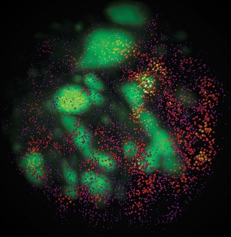
Showing 1-10 of 58 resultsTo image the accessible genome at nanometer scale in situ, we developed three-dimensional assay for transposase-accessible chromatin-photoactivated localization microscopy (3D ATAC-PALM) that integrates an assay for transposase-accessible chromatin with visualization, PALM super-resolution imaging and lattice light-sheet microscopy. Multiplexed with oligopaint DNA–fluorescence in situ hybridization (FISH), RNA–FISH and protein fluorescence, 3D ATAC-PALM connected microscopy and genomic data, revealing spatially segregated accessible chromatin domains (ACDs) that enclose active chromatin and transcribed genes. Using these methods to analyze genetically perturbed cells, we demonstrated that genome architectural protein CTCF prevents excessive clustering of accessible chromatin and decompacts ACDs. These results highlight 3D ATAC-PALM as a useful tool to probe the structure and organizing mechanism of the genome.
Combinatorial cis-regulatory networks encoded in animal genomes represent the foundational gene expression mechanism for directing cell-fate commitment and maintenance of cell identity by transcription factors (TFs). However, the 3D spatial organization of cis-elements and how such sub-nuclear structures influence TF activity remain poorly understood. Here, we combine lattice light-sheet imaging, single-molecule tracking, numerical simulations, and ChIP-exo mapping to localize and functionally probe Sox2 enhancer-organization in living embryonic stem cells. Sox2 enhancers form 3D-clusters that are segregated from heterochromatin but overlap with a subset of Pol II enriched regions. Sox2 searches for specific binding targets via a 3D-diffusion dominant mode when shuttling long-distances between clusters while chromatin-bound states predominate within individual clusters. Thus, enhancer clustering may reduce global search efficiency but enables rapid local fine-tuning of TF search parameters. Our results suggest an integrated model linking cis-element 3D spatial distribution to local-versus-global target search modalities essential for regulating eukaryotic gene transcription.
Few genetically dominant mutations involved in human disease have been fully explained at the molecular level. In cases where the mutant gene encodes a transcription factor, the dominant-negative mode of action of the mutant protein is particularly poorly understood. Here, we studied the genome-wide mechanism underlying a dominant-negative form of the SOX18 transcription factor (SOX18RaOp) responsible for both the classical mouse mutant Ragged Opossum and the human genetic disorder Hypotrichosis-lymphedema-telangiectasia-renal defect syndrome. Combining three single-molecule imaging assays in living cells together with genomics and proteomics analysis, we found that SOX18RaOp disrupts the system through an accumulation of molecular interferences which impair several functional properties of the wild-type SOX18 protein, including its target gene selection process. The dominant-negative effect is further amplified by poisoning the interactome of its wild-type counterpart, which perturbs regulatory nodes such as SOX7 and MEF2C. Our findings explain in unprecedented detail the multi-layered process that underpins the molecular aetiology of dominant-negative transcription factor function.
Transcription factor (TF)-directed enhanceosome assembly constitutes a fundamental regulatory mechanism driving spatiotemporal gene expression programs during animal development. Despite decades of study, we know little about the dynamics or order of events animating TF assembly at cis-regulatory elements in living cells and the long-range molecular "dialog" between enhancers and promoters. Here, combining genetic, genomic, and imaging approaches, we characterize a complex long-range enhancer cluster governing Krüppel-like factor 4 (Klf4) expression in naïve pluripotency. Genome editing by CRISPR/Cas9 revealed that OCT4 and SOX2 safeguard an accessible chromatin neighborhood to assist the binding of other TFs/cofactors to the enhancer. Single-molecule live-cell imaging uncovered that two naïve pluripotency TFs, STAT3 and ESRRB, interrogate chromatin in a highly dynamic manner, in which SOX2 promotes ESRRB target search and chromatin-binding dynamics through a direct protein-tethering mechanism. Together, our results support a highly dynamic yet intrinsically ordered enhanceosome assembly to maintain the finely balanced transcription program underlying naïve pluripotency.
Fluorescence microscopy relies on dyes that absorb and then emit photons. In addition to fluorescence, fluorophores can undergo photochemical processes that decrease quantum yield or result in spectral shifts and irreversible photobleaching. Chemical strategies that suppress these undesirable pathways—thereby increasing the brightness and photostability of fluorophores—are crucial for advancing the frontier of bioimaging. Here, we describe a general method to improve small-molecule fluorophores by incorporating deuterium into the alkylamino auxochromes of rhodamines and other dyes. This strategy increases fluorescence quantum yield, inhibits photochemically induced spectral shifts, and slows irreparable photobleaching, yielding next-generation labels with improved performance in cellular imaging experiments.
Expanding the palette of fluorescent dyes is vital to push the frontier of biological imaging. Although rhodamine dyes remain the premier type of small-molecule fluorophore owing to their bioavailability and brightness, variants excited with far-red or near-infrared light suffer from poor performance due to their propensity to adopt a lipophilic, nonfluorescent form. We report a framework for rationalizing rhodamine behavior in biological environments and a general chemical modification for rhodamines that optimizes long-wavelength variants and enables facile functionalization with different chemical groups. This strategy yields red-shifted 'Janelia Fluor' (JF) dyes useful for biological imaging experiments in cells and in vivo.
Neurons and glia operate in a highly coordinated fashion in the brain. Although glial cells have long been known to supply lipids to neurons via lipoprotein particles, new evidence reveals that lipid transport between neurons and glia is bidirectional. Here, we describe a co-culture system to study transfer of lipids and lipid-associated proteins from neurons to glia. The assay entails culturing neurons and glia on separate coverslips, pulsing the neurons with fluorescently labeled fatty acids, and then incubating the coverslips together. As astrocytes internalize and store neuron-derived fatty acids in lipid droplets, analyzing the number, size, and fluorescence intensity of lipid droplets containing the fluorescent fatty acids provides an easy and quantifiable measure of fatty acid transport. © 2019 The Authors.
TFIID-a complex of TATA-binding protein (TBP) and TBP-associated factors (TAFs)-is a central component of the Pol II promoter recognition apparatus. Recent studies have revealed significant downregulation of TFIID subunits in terminally differentiated myocytes, hepatocytes and adipocytes. Here, we report that TBP protein levels are tightly regulated by the ubiquitin-proteasome system. Using an in vitro ubiquitination assay coupled with biochemical fractionation, we identified Huwe1 as an E3 ligase targeting TBP for K48-linked ubiquitination and proteasome-mediated degradation. Upregulation of Huwe1 expression during myogenesis induces TBP degradation and myotube differentiation. We found that Huwe1 activity on TBP is antagonized by the deubiquitinase USP10, which protects TBP from degradation. Thus, modulating the levels of both Huwe1 and USP10 appears to fine-tune the requisite degradation of TBP during myogenesis. Together, our study unmasks a previously unknown interplay between an E3 ligase and a deubiquitinating enzyme regulating TBP levels during cellular differentiation.
Recent insights into genome organization have emphasized the importance of A/B chromatin compartments. While our previous research showed that Brd2 depletion weakens compartment boundaries and promotes A/B mixing 1, Hinojosa-Gonzalez et al.2 were unable to replicate the findings. In response, we revisited our Micro-C data and successfully replicated the original results using the default parameters in the cooltools software package. We show that, after correcting inconsistencies with the selection and phasing of the compartment profiles, the decrease in B compartment strength persists but the change in compartment identity is to a much lesser extent than originally reported. To further assess the regulatory role of Brd2, we used saddle plots to determine the strength of compartmentalization and observed a consistent decrease of compartment strength especially at B compartments upon Brd2 depletion. This study highlights the importance of selecting appropriate parameters and analytical tools for compartment analysis and carefully interpreting the results.
Liquid-liquid phase separation (LLPS) has emerged in recent years as an important physicochemical process for organizing diverse processes within cells via the formation of membraneless organelles termed biomolecular condensates. Emerging evidence now suggests that the formation and regulation of biomolecular condensates are also intricately linked to cancer formation and progression. We review the most recent literature linking the existence and/or dissolution of biomolecular condensates to different hallmarks of cancer formation and progression. We then discuss the opportunities that this condensate perspective provides for cancer research and the development of novel therapeutic approaches, including the perturbation of condensates by small-molecule inhibitors.