Filter
Associated Lab
- Remove Keller Lab filter Keller Lab
Publication Date
Type of Publication
3 Publications
Showing 1-3 of 3 results
The observation of biological processes in their natural in vivo context is a key requirement for quantitative experimental studies in the life sciences. In many instances, it will be crucial to achieve high temporal and spatial resolution over long periods of time without compromising the physiological development of the specimen. Here, we discuss the principles underlying light sheet-based fluorescence microscopes. The most recent implementation DSLM is a tool optimized to deliver quantitative data for entire embryos at high spatio-temporal resolution. We compare DSLM to the two established light microscopy techniques: confocal and two-photon fluorescence microscopy. DSLM provides up to 50 times higher imaging speeds and a 10-100 times higher signal-to-noise ratio, while exposing the specimens to at least three orders of magnitude less light energy than confocal and two-photon fluorescence microscopes. We conclude with a perspective for future development.
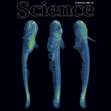
A long-standing goal of biology is to map the behavior of all cells during vertebrate embryogenesis. We developed digital scanned laser light sheet fluorescence microscopy and recorded nuclei localization and movement in entire wild-type and mutant zebrafish embryos over the first 24 hours of development. Multiview in vivo imaging at 1.5 billion voxels per minute provides "digital embryos," that is, comprehensive databases of cell positions, divisions, and migratory tracks. Our analysis of global cell division patterns reveals a maternally defined initial morphodynamic symmetry break, which identifies the embryonic body axis. We further derive a model of germ layer formation and show that the mesendoderm forms from one-third of the embryo’s cells in a single event. Our digital embryos, with 55 million nucleus entries, are provided as a resource.

Although microtubules are key players in many cellular processes, very little is known about their dynamic and mechanical properties in physiological three-dimensional environments. The conventional model of microtubule dynamic instability postulates two dynamic microtubule states, growth and shrinkage. However, several studies have indicated that such a model does not provide a comprehensive quantitative and qualitative description of microtubule behavior. Using three-dimensional laser light-sheet fluorescence microscopy and a three-dimensional sample preparation in spacious Teflon cylinders, we measured microtubule dynamic instability and elasticity in interphase Xenopus laevis egg extracts. Our data are inconsistent with a two-state model of microtubule dynamic instability and favor an extended four-state model with two independent metastable pause states over a three-state model with a single pause state. Moreover, our data on kinetic state transitions rule out a simple GTP cap model as the driving force of microtubule stabilization in egg extracts on timescales of a few seconds or longer. We determined the three-dimensional elastic properties of microtubules as a function of both the contour length and the dynamic state. Our results indicate that pausing microtubules are less flexible than growing microtubules and suggest a growth-speed-dependent persistence length. These data might hint toward mechanisms that enable microtubules to efficiently perform multiple different tasks in the cell and suggest the development of a unified model of microtubule dynamics and microtubule mechanics.