Filter
Associated Lab
- Ahrens Lab (2) Apply Ahrens Lab filter
- Baker Lab (1) Apply Baker Lab filter
- Druckmann Lab (3) Apply Druckmann Lab filter
- Harris Lab (3) Apply Harris Lab filter
- Hermundstad Lab (7) Apply Hermundstad Lab filter
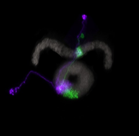
- Hess Lab (1) Apply Hess Lab filter
- Remove Jayaraman Lab filter Jayaraman Lab
- Ji Lab (1) Apply Ji Lab filter
- Karpova Lab (1) Apply Karpova Lab filter
- Looger Lab (10) Apply Looger Lab filter
- Podgorski Lab (1) Apply Podgorski Lab filter
- Reiser Lab (2) Apply Reiser Lab filter
- Romani Lab (4) Apply Romani Lab filter
- Rubin Lab (5) Apply Rubin Lab filter
- Saalfeld Lab (1) Apply Saalfeld Lab filter
- Scheffer Lab (1) Apply Scheffer Lab filter
- Schreiter Lab (9) Apply Schreiter Lab filter
- Svoboda Lab (9) Apply Svoboda Lab filter
- Zlatic Lab (1) Apply Zlatic Lab filter
Associated Project Team
Publication Date
- 2024 (1) Apply 2024 filter
- 2023 (1) Apply 2023 filter
- 2022 (3) Apply 2022 filter
- 2021 (1) Apply 2021 filter
- 2020 (4) Apply 2020 filter
- 2019 (4) Apply 2019 filter
- 2018 (3) Apply 2018 filter
- 2017 (4) Apply 2017 filter
- 2016 (3) Apply 2016 filter
- 2015 (4) Apply 2015 filter
- 2014 (1) Apply 2014 filter
- 2013 (3) Apply 2013 filter
- 2012 (2) Apply 2012 filter
- 2011 (2) Apply 2011 filter
- 2010 (2) Apply 2010 filter
- 2009 (2) Apply 2009 filter
- 2007 (1) Apply 2007 filter
- 2006 (1) Apply 2006 filter
- 2003 (1) Apply 2003 filter
Type of Publication
43 Publications
Showing 1-10 of 43 resultsThe neural circuits responsible for animal behavior remain largely unknown. We summarize new methods and present the circuitry of a large fraction of the brain of the fruit fly . Improved methods include new procedures to prepare, image, align, segment, find synapses in, and proofread such large data sets. We define cell types, refine computational compartments, and provide an exhaustive atlas of cell examples and types, many of them novel. We provide detailed circuits consisting of neurons and their chemical synapses for most of the central brain. We make the data public and simplify access, reducing the effort needed to answer circuit questions, and provide procedures linking the neurons defined by our analysis with genetic reagents. Biologically, we examine distributions of connection strengths, neural motifs on different scales, electrical consequences of compartmentalization, and evidence that maximizing packing density is an important criterion in the evolution of the fly's brain.
Flexible behaviors over long timescales are thought to engage recurrent neural networks in deep brain regions, which are experimentally challenging to study. In insects, recurrent circuit dynamics in a brain region called the central complex (CX) enable directed locomotion, sleep, and context- and experience-dependent spatial navigation. We describe the first complete electron-microscopy-based connectome of the CX, including all its neurons and circuits at synaptic resolution. We identified new CX neuron types, novel sensory and motor pathways, and network motifs that likely enable the CX to extract the fly's head-direction, maintain it with attractor dynamics, and combine it with other sensorimotor information to perform vector-based navigational computations. We also identified numerous pathways that may facilitate the selection of CX-driven behavioral patterns by context and internal state. The CX connectome provides a comprehensive blueprint necessary for a detailed understanding of network dynamics underlying sleep, flexible navigation, and state-dependent action selection.
Here we design and optimize a genetically encoded fluorescent indicator, iAChSnFR, for the ubiquitous neurotransmitter acetylcholine, based on a bacterial periplasmic binding protein. iAChSnFR shows large fluorescence changes, rapid rise and decay kinetics, and insensitivity to most cholinergic drugs. iAChSnFR revealed large transients in a variety of slice and in vivo preparations in mouse, fish, fly and worm. iAChSnFR will be useful for the study of acetylcholine in all animals.
Anchoring goals to spatial representations enables flexible navigation in both animals and artificial agents. However, using this strategy can be challenging in novel environments, when both spatial and goal representations must be acquired quickly and simultaneously. Here, we propose a framework for how Drosophila use their internal representation of head direction to build a goal heading representation upon selective thermal reinforcement. We show that flies in a well-established operant visual learning paradigm use stochastically generated fixations and directed saccades to express heading preferences, and that compass neurons, which represent flies’ head direction, are required to modify these preferences based on reinforcement. We describe how flies’ ability to quickly map their surroundings and adapt their behavior to the rules of their environment may rest on a behavioral policy whose parameters are flexible but whose form and dependence on head direction and goal representations are genetically encoded in the modular structure of their circuits. Using a symmetric visual setting, which predictably alters the dynamics of the head direction system, enabled us to describe how interactions between the evolving representations of head direction and goal impact behavior. We show how a policy tethered to these two internal representations can facilitate rapid learning of new goal headings, drive more exploitative behavior about stronger goal headings, and ensure that separate learning processes involved in mapping the environment and forming goals within that environment remain consistent with one another. Many of the mechanisms we outline may be broadly relevant for rapidly adaptive behavior driven by internal representations.
Fluorescent protein-based sensors for detecting neuronal activity have been developed largely based on non-neuronal screening systems. However, the dynamics of neuronal state variables (e.g., voltage, calcium, etc.) are typically very rapid compared to those of non-excitable cells. We developed an electrical stimulation and fluorescence imaging platform based on dissociated rat primary neuronal cultures. We describe its use in testing genetically-encoded calcium indicators (GECIs). Efficient neuronal GECI expression was achieved using lentiviruses containing a neuronal-selective gene promoter. Action potentials (APs) and thus neuronal calcium levels were quantitatively controlled by electrical field stimulation, and fluorescence images were recorded. Images were segmented to extract fluorescence signals corresponding to individual GECI-expressing neurons, which improved sensitivity over full-field measurements. We demonstrate the superiority of screening GECIs in neurons compared with solution measurements. Neuronal screening was useful for efficient identification of variants with both improved response kinetics and high signal amplitudes. This platform can be used to screen many types of sensors with cellular resolution under realistic conditions where neuronal state variables are in relevant ranges with respect to timing and amplitude.
Navigating animals continuously integrate velocity signals to update internal representations of their directional heading and spatial location in the environment. How neural circuits combine sensory and motor information to construct these velocity estimates and how these self-motion signals, in turn, update internal representations that support navigational computations are not well understood. Recent work in Drosophila has identified a neural circuit that performs angular path integration to compute the fly's head direction, but the nature of the velocity signal is unknown. Here we identify a pair of neurons necessary for angular path integration that encode the fly's rotational velocity with high accuracy using both visual optic flow and motor information. This estimate of rotational velocity does not rely on a moment-to-moment integration of sensory and motor information. Rather, when visual and motor signals are congruent, these neurons prioritize motor information over visual information, and when the two signals are in conflict, reciprocal inhibition selects either the motor or visual signal. Together, our results suggest that flies update their head direction representation by constructing an estimate of rotational velocity that relies primarily on motor information and only incorporates optic flow signals in specific sensorimotor contexts, such as when the motor signal is absent.
To flexibly navigate, many animals rely on internal spatial representations that persist when the animal is standing still in darkness, and update accurately by integrating the animal's movements in the absence of localizing sensory cues. Theories of mammalian head direction cells have proposed that these dynamics can be realized in a special class of networks that maintain a localized bump of activity via structured recurrent connectivity, and that shift this bump of activity via angular velocity input. Although there are many different variants of these so-called ring attractor networks, they all rely on large numbers of neurons to generate representations that persist in the absence of input and accurately integrate angular velocity input. Surprisingly, in the fly, Drosophila melanogaster, a head direction representation is maintained by a much smaller number of neurons whose dynamics and connectivity resemble those of a ring attractor network. These findings challenge our understanding of ring attractors and their putative implementation in neural circuits. Here, we analyzed failures of angular velocity integration that emerge in small attractor networks with only a few computational units. Motivated by the peak performance of the fly head direction system in darkness, we mathematically derived conditions under which small networks, even with as few as 4 neurons, achieve the performance of much larger networks. The resulting description reveals that by appropriately tuning the network connectivity, the network can maintain persistent representations over the continuum of head directions, and it can accurately integrate angular velocity inputs. We then analytically determined how performance degrades as the connectivity deviates from this optimally-tuned setting, and we find a trade-off between network size and the tuning precision needed to achieve persistence and accurate integration. This work shows how even small networks can accurately track an animal's movements to guide navigation, and it informs our understanding of the functional capabilities of discrete systems more broadly.
Many animals maintain an internal representation of their heading as they move through their surroundings. Such a compass representation was recently discovered in a neural population in the Drosophila melanogaster central complex, a brain region implicated in spatial navigation. Here, we use two-photon calcium imaging and electrophysiology in head-fixed walking flies to identify a different neural population that conjunctively encodes heading and angular velocity, and is excited selectively by turns in either the clockwise or counterclockwise direction. We show how these mirror-symmetric turn responses combine with the neurons' connectivity to the compass neurons to create an elegant mechanism for updating the fly's heading representation when the animal turns in darkness. This mechanism, which employs recurrent loops with an angular shift, bears a resemblance to those proposed in theoretical models for rodent head direction cells. Our results provide a striking example of structure matching function for a broadly relevant computation.
The central complex is a highly conserved insect brain region composed of morphologically stereotyped neurons that arborize in distinctively shaped substructures. The region is implicated in a wide range of behaviors and several modeling studies have explored its circuit computations. Most studies have relied on assumptions about connectivity between neurons based on their overlap in light microscopy images. Here, we present an extensive functional connectome of Drosophila melanogaster's central complex at cell-type resolution. Using simultaneous optogenetic stimulation, calcium imaging and pharmacology, we tested the connectivity between 70 presynaptic-to-postsynaptic cell-type pairs. We identi1ed numerous inputs to the central complex, but only a small number of output channels. Additionally, the connectivity of this highly recurrent circuit appears to be sparser than anticipated from light microscopy images. Finally, the connectivity matrix highlights the potentially critical role of a class of bottleneck interneurons. All data is provided for interactive exploration on a website.
Clock neurons generate circadian rhythms in behavioral activity, but the relevant pathways remain poorly understood. In this issue of Neuron, Liang et al. (2019) show that distinct clock neurons independently drive movement-promoting “ring neurons” in Drosophila through dopaminergic relays to support morning and evening locomotor activity.
View Publication Page