Filter
Associated Lab
Associated Project Team
Publication Date
- 2015 (2) Apply 2015 filter
- 2014 (1) Apply 2014 filter
- 2011 (3) Apply 2011 filter
- 2010 (1) Apply 2010 filter
- 2009 (2) Apply 2009 filter
- 2008 (1) Apply 2008 filter
- 2006 (3) Apply 2006 filter
- 2005 (1) Apply 2005 filter
- 2004 (1) Apply 2004 filter
- 2003 (2) Apply 2003 filter
- 2002 (1) Apply 2002 filter
- 2001 (3) Apply 2001 filter
- 2000 (3) Apply 2000 filter
- 1985 (1) Apply 1985 filter
Type of Publication
25 Publications
Showing 1-10 of 25 resultsA novel family of candidate gustatory receptors (GRs) was recently identified in searches of the Drosophila genome. We have performed in situ hybridization and transgene experiments that reveal expression of these genes in both gustatory and olfactory neurons in adult flies and larvae. This gene family is likely to encode both odorant and taste receptors. We have visualized the projections of chemosensory neurons in the larval brain and observe that neurons expressing different GRs project to discrete loci in the antennal lobe and subesophageal ganglion. These data provide insight into the diversity of chemosensory recognition and an initial view of the representation of gustatory information in the fly brain.
The taste system is one of our fundamental senses, responsible for detecting and responding to sweet, bitter, umami, salty, and sour stimuli. In the tongue, the five basic tastes are mediated by separate classes of taste receptor cells each finely tuned to a single taste quality. We explored the logic of taste coding in the brain by examining how sweet, bitter, umami, and salty qualities are represented in the primary taste cortex of mice. We used in vivo two-photon calcium imaging to demonstrate topographic segregation in the functional architecture of the gustatory cortex. Each taste quality is represented in its own separate cortical field, revealing the existence of a gustotopic map in the brain. These results expose the basic logic for the central representation of taste.
Light-induced photoreceptor apoptosis occurs in many forms of inherited retinal degeneration resulting in blindness in both vertebrates and invertebrates. Though mutations in several photoreceptor signaling proteins have been implicated in triggering this process, the molecular events relating light activation of rhodopsin to photoreceptor death are yet unclear. Here, we uncover a pathway by which activation of rhodopsin in Drosophila mediates apoptosis through a G protein-independent mechanism. This process involves the formation of membrane complexes of phosphorylated, activated rhodopsin and its inhibitory protein arrestin, and subsequent clathrin-dependent endocytosis of these complexes into a cytoplasmic compartment. Together, these data define the proapoptotic molecules in Drosophila photoreceptors and indicate a novel signaling pathway for light-activated rhodopsin molecules in control of photoreceptor viability.
In mammals, taste perception is a major mode of sensory input. We have identified a novel family of 40-80 human and rodent G protein-coupled receptors expressed in subsets of taste receptor cells of the tongue and palate epithelia. These candidate taste receptors (T2Rs) are organized in the genome in clusters and are genetically linked to loci that influence bitter perception in mice and humans. Notably, a single taste receptor cell expresses a large repertoire of T2Rs, suggesting that each cell may be capable of recognizing multiple tastants. T2Rs are exclusively expressed in taste receptor cells that contain the G protein alpha subunit gustducin, implying that they function as gustducin-linked receptors. In the accompanying paper, we demonstrate that T2Rs couple to gustducin in vitro, and respond to bitter tastants in a functional expression assay.
The sense of taste provides animals with valuable information about the nature and quality of food. Mammals can recognize and respond to a diverse repertoire of chemical entities, including sugars, salts, acids and a wide range of toxic substances. Several amino acids taste sweet or delicious (umami) to humans, and are attractive to rodents and other animals. This is noteworthy because L-amino acids function as the building blocks of proteins, as biosynthetic precursors of many biologically relevant small molecules, and as metabolic fuel. Thus, having a taste pathway dedicated to their detection probably had significant evolutionary implications. Here we identify and characterize a mammalian amino-acid taste receptor. This receptor, T1R1+3, is a heteromer of the taste-specific T1R1 and T1R3 G-protein-coupled receptors. We demonstrate that T1R1 and T1R3 combine to function as a broadly tuned L-amino-acid sensor responding to most of the 20 standard amino acids, but not to their D-enantiomers or other compounds. We also show that sequence differences in T1R receptors within and between species (human and mouse) can significantly influence the selectivity and specificity of taste responses.
Mammals can taste a wide repertoire of chemosensory stimuli. Two unrelated families of receptors (T1Rs and T2Rs) mediate responses to sweet, amino acids, and bitter compounds. Here, we demonstrate that knockouts of TRPM5, a taste TRP ion channel, or PLCbeta2, a phospholipase C selectively expressed in taste tissue, abolish sweet, amino acid, and bitter taste reception, but do not impact sour or salty tastes. Therefore, despite relying on different receptors, sweet, amino acid, and bitter transduction converge on common signaling molecules. Using PLCbeta2 taste-blind animals, we then examined a fundamental question in taste perception: how taste modalities are encoded at the cellular level. Mice engineered to rescue PLCbeta2 function exclusively in bitter-receptor expressing cells respond normally to bitter tastants but do not taste sweet or amino acid stimuli. Thus, bitter is encoded independently of sweet and amino acids, and taste receptor cells are not broadly tuned across these modalities.
The sense of taste is a specialized chemosensory system dedicated to the evaluation of food and drink. Despite the fact that vertebrates and insects have independently evolved distinct anatomic and molecular pathways for taste sensation, there are clear parallels in the organization and coding logic between the two systems. There is now persuasive evidence that tastant quality is mediated by labeled lines, whereby distinct and strictly segregated populations of taste receptor cells encode each of the taste qualities.
The evolution of the ancestral eukaryotic flagellum is an example of a cellular organelle that became dispensable in some modern eukaryotes while remaining an essential motile and sensory apparatus in others. To help define the repertoire of specialized proteins needed for the formation and function of cilia, we used comparative genomics to analyze the genomes of organisms with prototypical cilia, modified cilia, or no cilia and identified approximately 200 genes that are absent in the genomes of nonciliated eukaryotes but are conserved in ciliated organisms. Importantly, over 80% of the known ancestral proteins involved in cilia function are included in this small collection. Using Drosophila as a model system, we then characterized a novel family of proteins (OSEGs: outer segment) essential for ciliogenesis. We show that osegs encode components of a specialized transport pathway unique to the cilia compartment and are related to prototypical intracellular transport proteins.
In Drosophila photoreceptors the multivalent PDZ protein INAD organizes the phototransduction cascade into a macromolecular signaling complex containing the effector PLC, the light-activated TRP channels, and a regulatory PKC. Previously, we showed that the subcellular localization of INAD signaling complexes is critical for signaling. Now we have examined how INAD complexes are anchored and assembled in photoreceptor cells. We find that trp mutants, or transgenic flies expressing inaD alleles that disrupt the interaction between INAD and TRP, cause the mislocalization of the entire transduction complex. The INAD-TRP interaction is not required for targeting but rather for anchoring of complexes, because INAD and TRP can be targeted independently of each other. We also show that, in addition to its scaffold role, INAD functions to preassemble transduction complexes. Preassembly of signaling complexes helps to ensure that transduction complexes with the appropriate composition end up in the proper location. This may be a general mechanism used by cells to target different signaling machinery to the pertinent subcellular location.
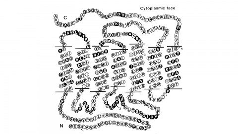
Using a novel method for detecting cross-homologous nucleic acid sequences we have isolated the gene coding for the major rhodopsin of Drosophila melanogaster and mapped it to chromosomal region 92B8-11. Comparison of cDNA and genomic DNA sequences indicates that the gene is divided into five exons. The amino acid sequence deduced from the nucleotide sequence is 373 residues long, and the polypeptide chain contains seven hydrophobic segments that appear to correspond to the seven transmembrane segments characteristic of other rhodopsins. Three regions of Drosophila rhodopsin are highly conserved with the corresponding domains of bovine rhodopsin, suggesting an important role for these polypeptide regions.